PPT Anatomy of the Male Reproductive System PowerPoint Presentation, free download ID883757
Within the seminiferous tubules, somatic Sertoli cells extend from the basement membrane adjacent to PTM cells to the lumen and surround the developing germ cells (Figure 16.1(C)).Germ cells are present throughout the seminiferous tubule epithelium, with the most immature and undifferentiated cells, including spermatogonial stem cells (SSCs), present on the basement membrane and the most.
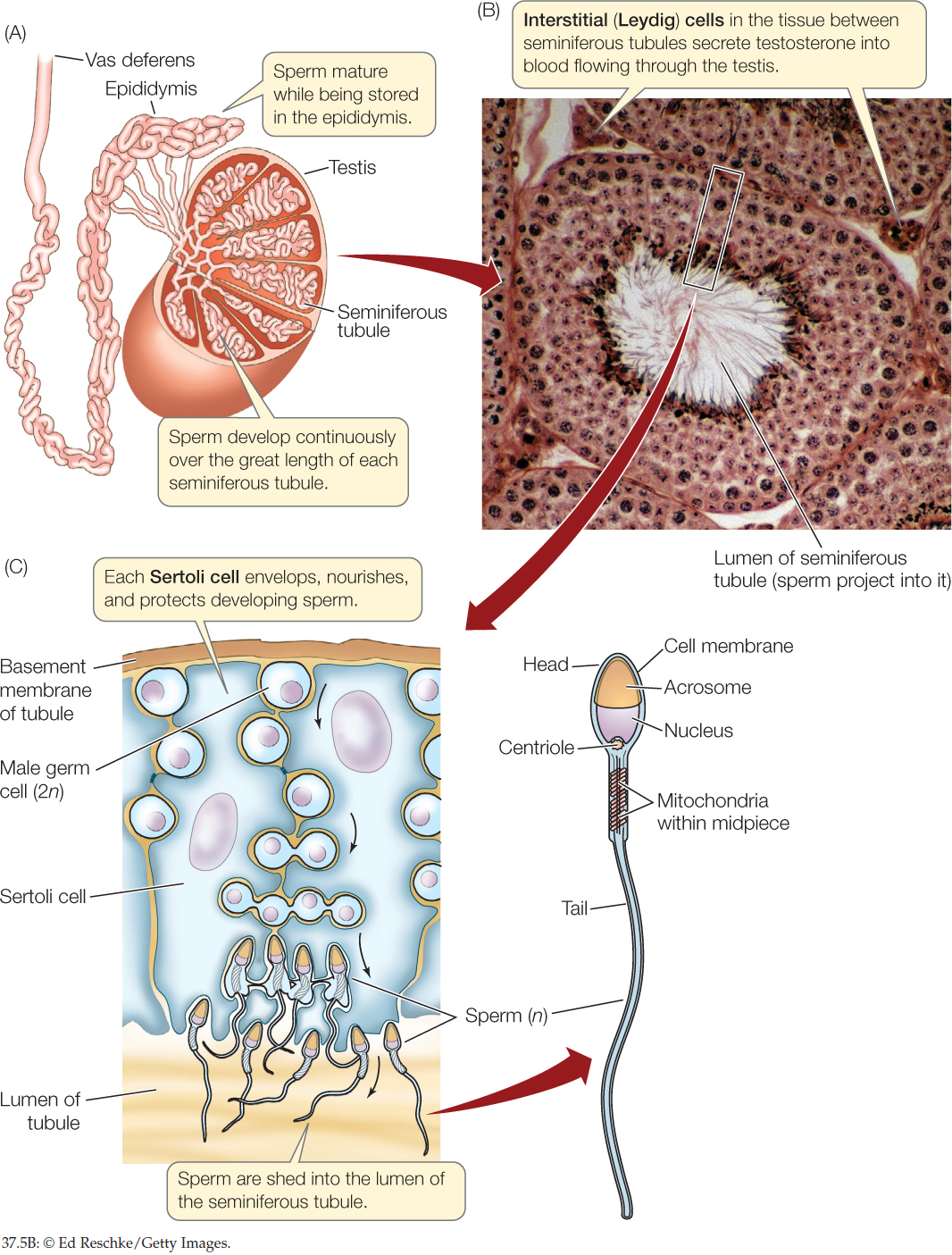
Figure 37.5
An important term in the title 'seminiferous tubule' is the word seminiferous, which means bearing a seed or producing sperm in the context of biology. This meaning gives a hint on what.

Male Reproductive System Lab
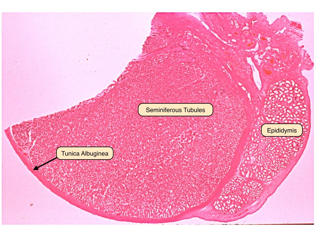
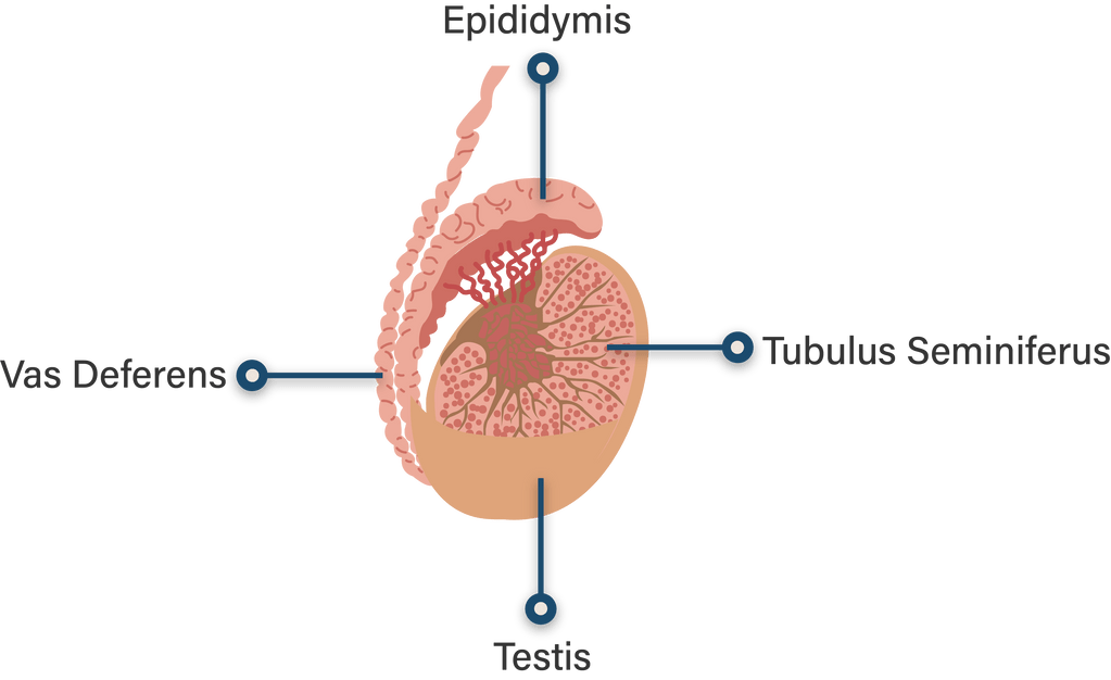
Other articles where seminiferous tubule is discussed: animal reproductive system: Testes:.testes are composed largely of seminiferous tubules—coiled tubes, the walls of which contain cells that produce sperm—and are surrounded by a capsule, the tunica albuginea. Seminiferous tubules may constitute up to 90 percent of the testis. The tubule walls consist of a multilayered germinal.
Seminiferous Tubules
One of the major concerns of the world health community is the infertility. The definition of infertility according to the World Health Organization (WHO) and the American Society for Reproductive Medicine (ASRM) is the inability of a healthy couple to achieve a conception after one year of regular, unprotected intercourse. Fertility complications affect seven percent of the male. The causes.
Male Seminiferous Tubules
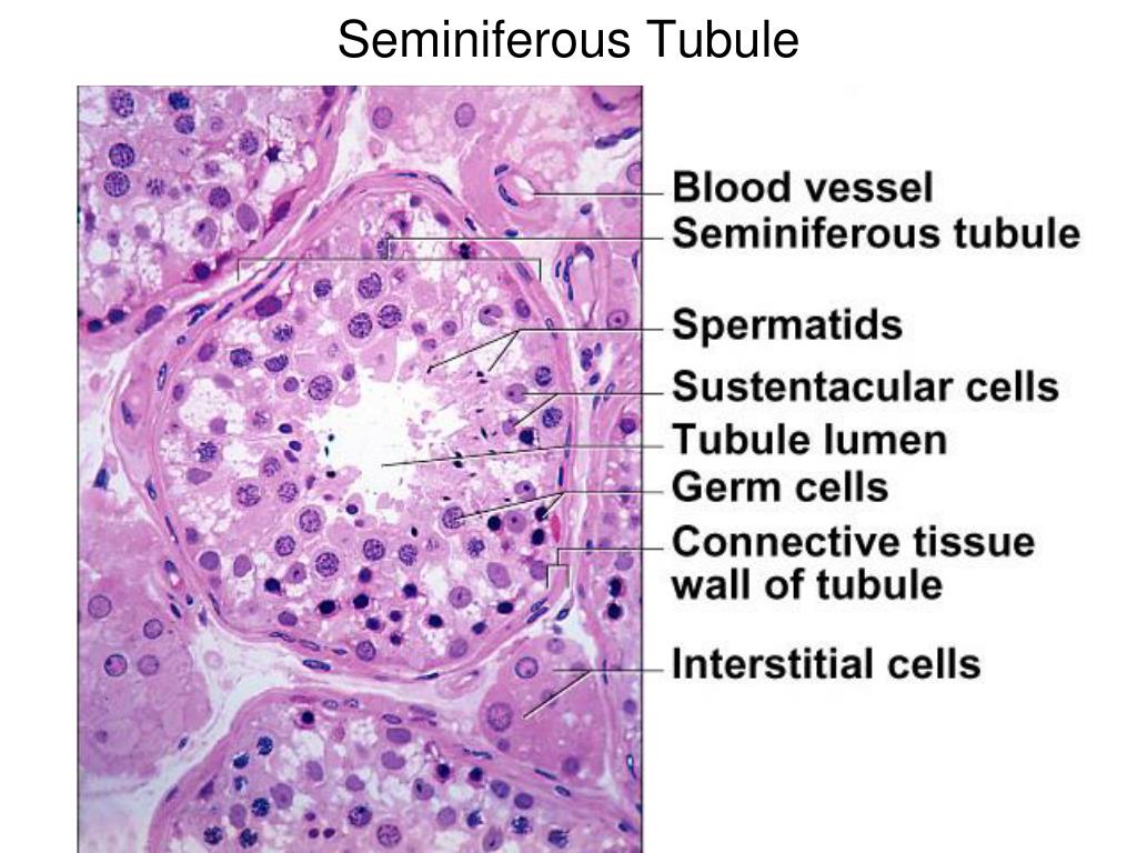
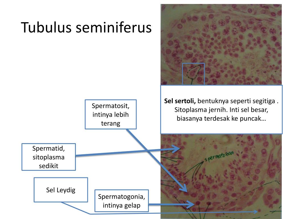
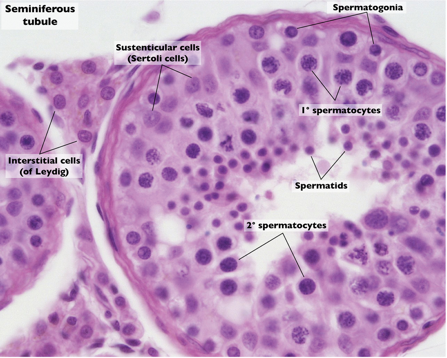
Reproductive - Histology of seminiferous tubules View Related Images. Description: This image is a histological section of the seminiferous tubules.
jelaskan apa itu tubulus seminiferus...
Junctional complexes are formed between the adjacent Sertoli cells (Sertoli-Sertoli barrier or blood-testis barrier) and this barrier separate the space within the seminiferous tubule into basal and luminal compartments. A layer of peritubular myoid cells surround the tubules. The nuclei of Sertoli cells are circled and nuclei of spermatogonia are enclosed by a rectangle.

Post Medizzystructure Of Testis learnbrainly.live
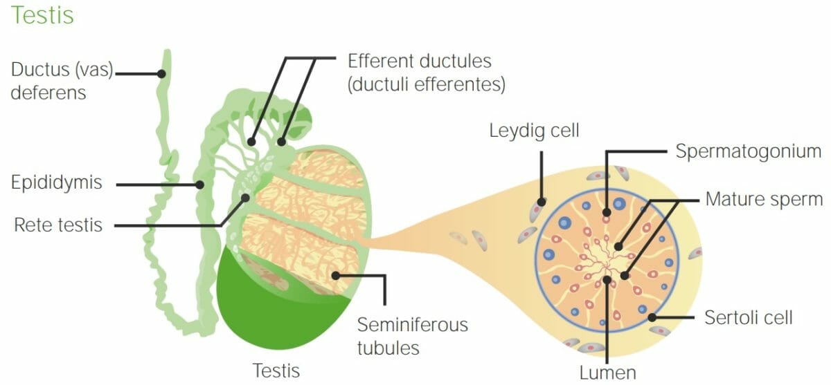
The testes. Seminiferous tubules comprise the bulk (80%) of the testis mass and are responsible for developing germ cells. Support cells include Sertoli cells, fibrocyte, and myoid cells. The interstitium between the seminiferous tubules contains blood vessels, lymphatics, Leydig cells, mast cells, nerves.

Gambaran Histologi Tubulus Seminiferus Dari Sediaan Testis Mencit
A simple structure is observed uniformly throughout the entire length of the ~ 2 m (!) long seminiferous tubules, which is tightly packed within a single mouse testis (Fig. 3) (Hilton & Turner, 1993; Russell et al., 1990; Yoshida, 2018a).Sertoli cells and two layers of peritubular myoid and lymphatic endothelial cells along with a basement membrane compose the basic structural framework of.

Histology of seminiferous tubule of testis YouTube
Seminiferous tubules develop from sex cords, which are embryonic structures with simple C-shaped arches. Histologically, the epithelium of adult mouse seminiferous tubules has been divided into 12 stages based on the associations of spermatogenic cells in four cycles of spermatogenesis. However, the gross characteristics of the seminiferous.
PPT Organ Reproduksi Pria PowerPoint Presentation, free download ID4739934
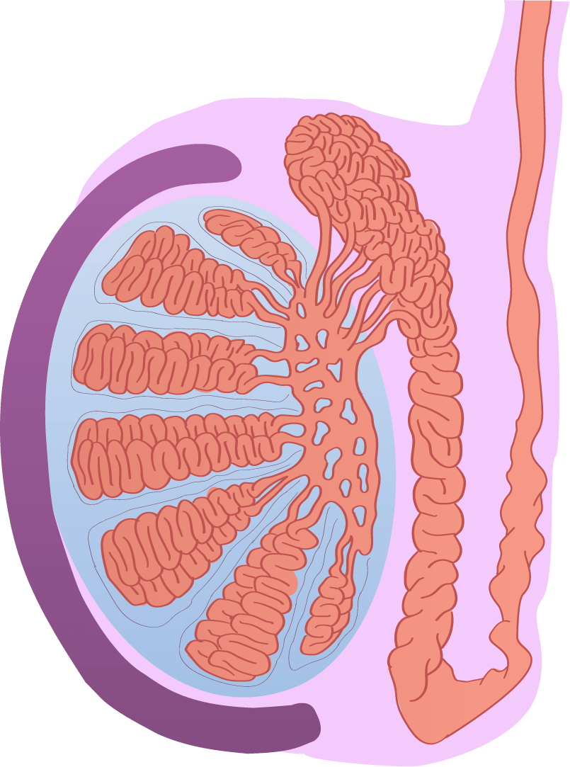
Definition. Each lobule of testis is contained in one of the intervals between the fibrous septa which extend between the mediastinum testis and the tunica albuginea, and consists of from one to three, or more, minute convoluted tubes, the convoluted seminiferous tubules.
jelaskan apa itu tubulus seminiferus...
Seminiferous tubules have a highly simple and uniform structure over their total lengths of up to 2 m coiled within the mouse testis ( Hilton & Turner, 1993; Russell et al., 1990; Yoshida, 2018a ). SSCs are scattered over the basal compartment with strikingly constant average densities ( Kitadate et al., 2019 ), making it simple to obtain large.

The Male Reproductive System Boundless Anatomy and Physiology
We developed a technique to analyze the high-resolution three-dimensional (3D) structure of seminiferous tubules. It consists of segmentation of tubules in serial paraffin sections of the testis by marking the basement membrane with periodic acid-Schiff or a fluorescent anti-laminin antibody followed by 3D reconstruction of tubules with high-performance software. Using this method, we.

PPT MALE REPRODUCTIVE SYSTEM PowerPoint Presentation, free download ID2000162
The seminiferous tubules are the site of the germination, maturation, and transportation of the sperm cells within the male testes. Seminiferous tubules are made up of columnar Sertoli cells surrounded by spermatogenic cells on the epithelial interior and stem cells exteriorly. Spermatogenesis through the process of meiosis takes place within.

Struktur, Bagian, dan Fungsi Alat Reproduksi LakiLaki Materi Sistem Reproduksi Biologi
Fungsi dari Tubulus Seminiferous. Tanpa adanya sel sperma, manusia serta hewan tentu tidak dapat melakukan reproduksi generatif dengan berhasil. Sperma tersebut dihasilkan oleh tubulus Seminiferus. Selain menghasilkan sel sperma, tubulus Seminiferus juga memiliki fungsi lain seperti menjaga sel sperma, serta untuk menyimpan sperma.
Gonadal Hormones Concise Medical Knowledge
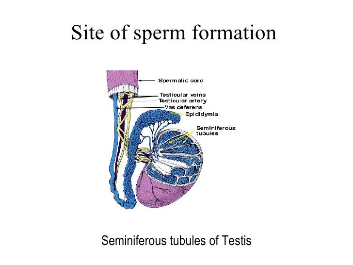
Abstract. The testis is made up of the seminiferous tubules, where the spermatozoa develop, and interstitial cells (Leydig cells), where testosterone is produced. The seminiferous tubules are long U-shaped tubules which form their U-turn at the periphery of the testis as both distal ends drain toward the central superior and posterior regions.
Human Testicles Anatomy, Inside Structure, Function and Location
During the third [ (, or condensing spermatid)] stage, the [spermatids] become fully developed. The filament for the formation of the midpiece [ (of the flagellum, or tail)] connects to the head. The cellular protoplasm gradually disappears, generating the well-known appendage joined to the midpiece.