Sinus XRay Positioning An XRay Guide Medical Professionals
Mucoceles of the paranasal accessory sinuses are relatively uncommon lesions which, though their etiology is still controversial and incompletely understood, are generally attributed to some form of local obstruction. Although histologically benign and slow in growth, they are prone to result in facial and ocular deformities and may on occasion produce alarming, if not ultimately serious.

Paranasal sinuses, head Xrays Stock Image C023/8535 Science Photo Library
Clinical Relevance: Sinusitis. As the paranasal sinuses are continuous with the nasal cavity, an upper respiratory tract infection can spread to the sinuses. Infection of the sinuses causes inflammation (particularly pain and swelling) of the mucosa, and is known as sinusitis. If more than one sinus is affected, it is called pansinusitis.
Radiografia dei seni paranasali (proiezione frontale) DocCheck
The Paranasal Sinuses: Normal Roentgen Anatomy By LEWIS E. ETTER, M.D. THE PARANASAL SINUSES consist of the ethmoid cells and the maxil- lary, frontal and sphenoid sinuses. Thev are air-containing adjuncts to the respiratory system which communicate with the nasal cavities and serve principally to warm and humidify the inspired air. They also.
PARANASAL SINUSES Radiology Key
The keynote of this presentation may be summarized in the terse question, "Is the referring physician getting sufficient and accurate information from the average roentgen examination of the paranasal sinuses?" I believe not. I frequently review and confer on films made elsewhere, and find the majority of them to be sadly deficient in many.
X Ray Of The Paranasal Sinuses Bilateral Maxillary Sinusitis Stock My XXX Hot Girl
Background: The authors reviewed treatment results in patients with nasal and paranasal sinus carcinoma from a large retrospective cohort and conducted a systematic literature review. Methods: Two hundred twenty patients who were treated between 1975 and 1994 with a minimum follow-up of 4 years were reviewed retrospectively. A systematic review of published articles on patients with.

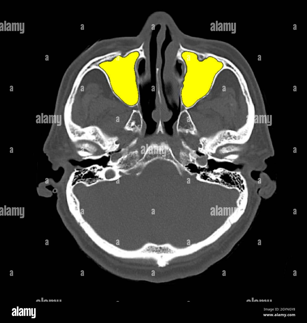
CT scan of the paranasal sinuses with contrast. The coronal and axial... Download Scientific
dr. Meliyana. Rontgen sinus digunakan untuk menilai kelainan struktur anatomi sinus paranasal, seperti sinusitis, polip, dan fraktur. Kelainan pada sinus paranasal yang dapat dinilai melalui pemeriksaan rontgen sinus adalah anomali kongenital, tumor, inflamasi, kondisi alergi, komplikasi dari infeksi, obstruksi, dan trauma. [1,2]
Sinuses Radiographic Anatomy wikiRadiography
1. Fifty-one per cent of patients with suspected sinusitis have abnormal roentgenograms consisting of one alone or a combination of mucosal thickening, opacity, fluid and polyps. 2. Similar abnormal roentgenograms are found in 57 per cent of children without sinusitis and in 75 per cent of children who have an upper respiratory infection, but without sinusitis. 3. Only 6 of 120 patients with.
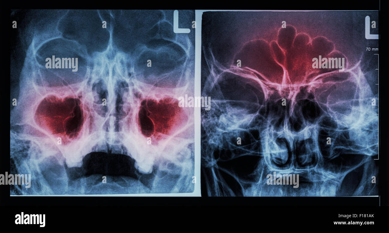
Film Xray paranasal sinus show sinusitis at maxillary sinus ( left image ) , frontal sinus
The widespread employment of roentgen methods in the examination of paranasal sinus disease suggests that these methods are of considerable practical value. In an effort to determine to what extent this assumption is correct, roentgenologic and clinical observations regarding the sinuses have been reviewed in a considerable number of case records. Roentgenologic impressions have been compared.

INTERPRETASI RADIOLOGI SINUS PARANASAL YouTube
During the meeting of this section at San Francisco in 1923 it was my privilege to describe 1 in detail two positions for making roentgenograms of the paranasal sinuses, more especially the sphenoids and ethmoids. I adopted these after painstaking experiments with dried skulls which had the sphenoids and two groups of ethmoids filled with substances of different ray-absorbing values, because.

Coronal computed tomogram through the paranasal sinuses The BMJ
The nasal cavity is a roughly cylindrical, midline airway passage that extends from the nasal ala anteriorly to the choana posteriorly.[1] It is divided in the midline by the nasal septum. On each side, it is flanked by the maxillary sinuses and roofed by the frontal, ethmoid, and sphenoid sinuses in an anterior to posterior fashion.[1] While seemingly simple, sinonasal anatomy is composed of.

CT scan of paranasal sinus — axial view showing inward retraction of... Download Scientific
The paranasal sinuses (the hollow spaces in the skull and facial bones around the nose) are air-filled cavities within the frontal, ethmoidal, sphenoidal, and maxillary bones.[1] They are outgrowths from the nasal cavity. All of them drain into the superior or lateral aspect of the nose.[2] The sinuses' lining mucosa is continuous with the nasal cavity; therefore, any infections from the nasal.

コンプリート! paranasal sinuses x ray labeled 481856Paranasal sinuses x ray anatomy
The rontgen ray examination of the paranasal sinuses with particular reference to the frontal sinuses.. The rontgen ray examination of the paranasal sinuses with particular reference to the frontal sinuses Br J Radiol. 1948 Sep;21(249):431-7. doi: 10.1259/0007-1285-21-249-431. Author S WELIN. PMID:.
Sinus XRay Positioning An XRay Guide Medical Professionals
The frontal sinuses are paired triangular-shaped cavities located in the frontal bones. They are the most superior paranasal sinuses, situated deep to the superciliary arches and the root of the nose. The frontal sinuses are drained via the frontonasal duct to the ethmoidal infundibulum.This infundibulum then opens into the middle nasal meatus via the semilunar hiatus.
Paranasal sinuses hires stock photography and images Alamy
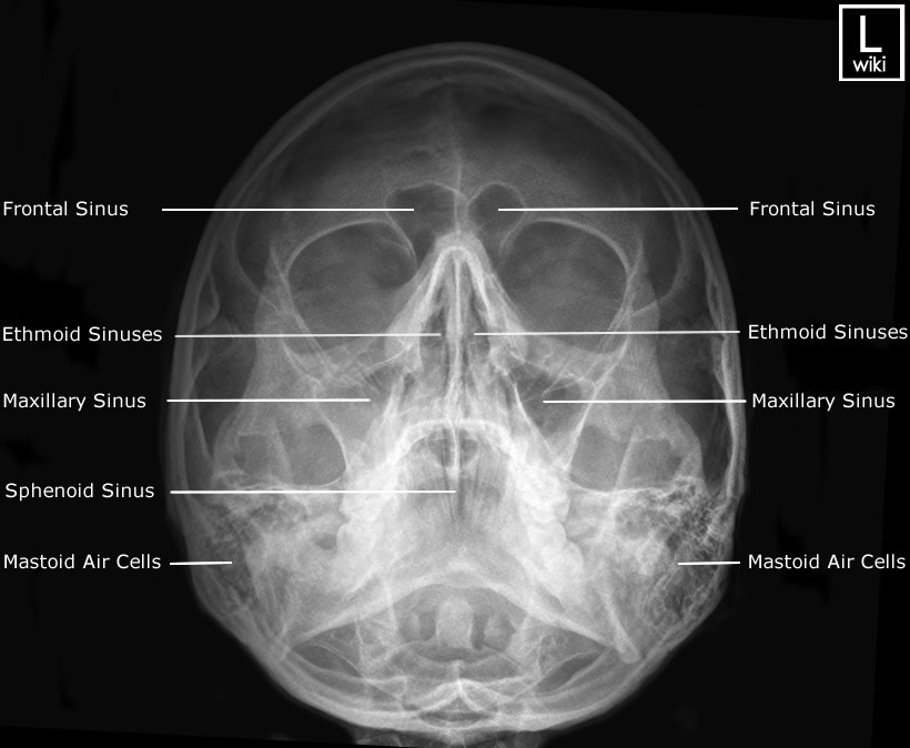
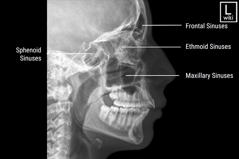
Sinus Series. What you need to know about…. The paranasal sinuses are a group of air-filled cavities located in the facial area. The maxillary sinuses are located under each of the eyes, the frontal sinus is located in the area of the forehead directly above the nose, the ethmoidal sinuses are located in the area of the eyes and the upper.

Image
Some of the anatomic variants have been reported to be associated with chronic rhinosinusitis, possibly leading to inflammation by obstructing drainage pathways from the sinuses and nasal cavity [2-5, 10].Specifically, large ethmoidal bullae correlated with maxillary sinusitis in one study [], but another study [] showed a correlation between paradoxically bent middle turbinates.
Paranasal sinus anatomy, CT scan Stock Photo Alamy
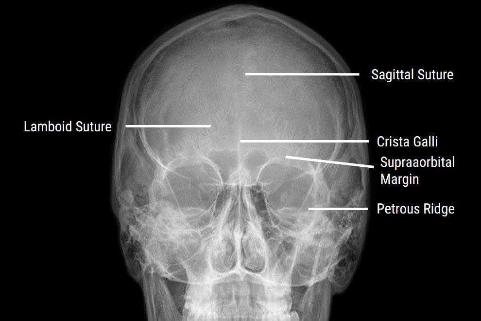
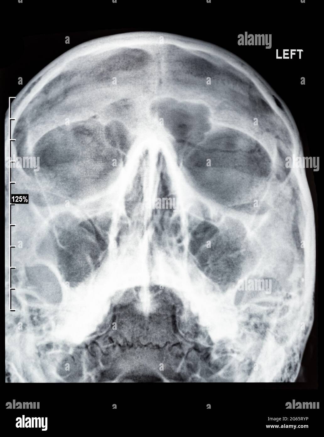
This view aids in visualizing the paranasal sinuses, especially the frontal sinus 4. It can help to assess inflammatory conditions such as sinusitis and secondary osteomyelitis, and sinus polyps or cysts. Additionally, skull fractures, neoplastic processes, or Paget disease may also be visualized via this view 4. Patient position