Human Skeleton Bones Labeled Image Anatomy System Human Body
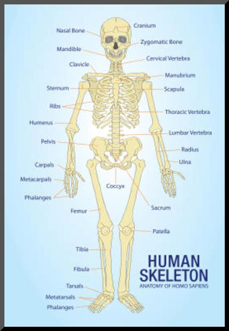
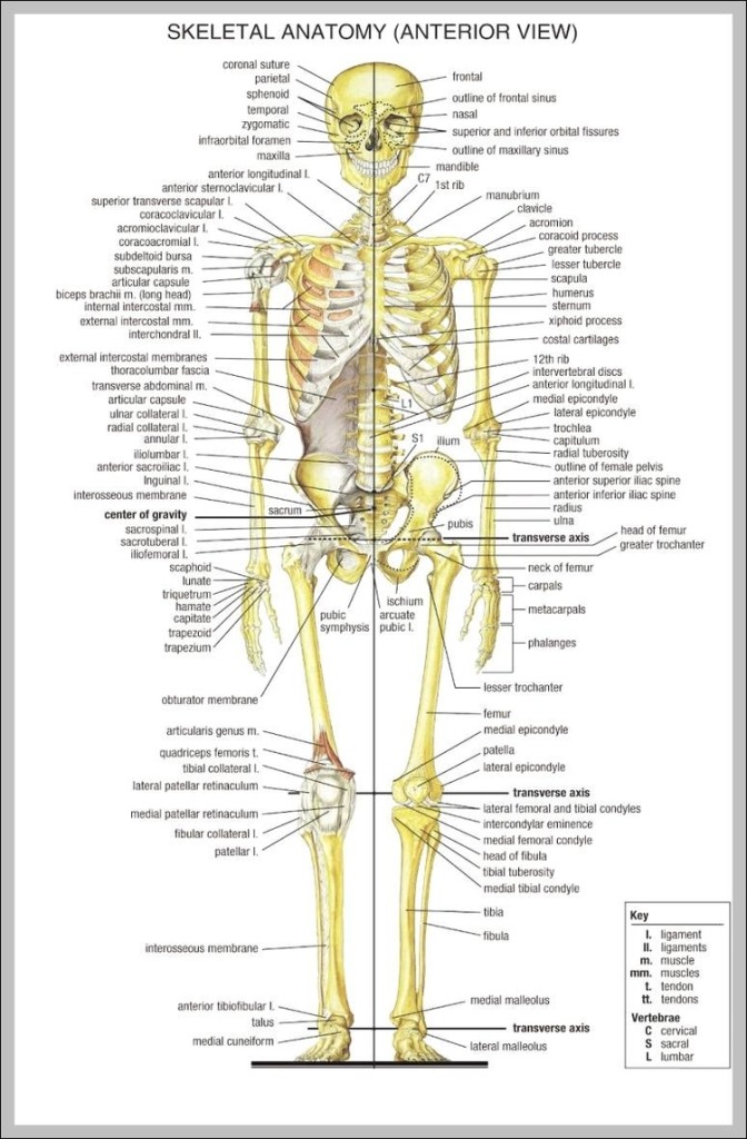
Figure \(\PageIndex{2}\): Some of the 206 bones are labeled on the adult human skeleton. Besides bones, the skeletal system includes cartilage and ligaments. Cartilage is a type of dense connective tissue, made of tough protein fibers. It is strong but flexible and very smooth. It covers the ends of bones at joints, providing a smooth surface.
Bone anatomy labeled diagram hires stock photography and images Alamy
The human skeleton is the internal framework of the human body. It is composed of around 270 bones at birth - this total decreases to around 206 bones by adulthood after some bones get fused together. [1] The bone mass in the skeleton makes up about 14% of the total body weight (ca. 10-11 kg for an average person) and reaches maximum mass.
human bones labelled diagram
Summary. The skeletal system is made up of your bones, ligaments, and cartilage. Though its main function is to provide structural support for the body, it also stores important minerals—such as calcium—forms red blood cells, and protects your internal organs. The skeletal system can break down into two main categories—the axial skeleton.
Skeleton Bones Labeled
The cranium is a skull bone that covers the brain, as seen in the skeleton diagram. The facial bones are not a part of the cranium. The bones that are just above the ear or in front of the ear are known as temporal bones. Stapes. Stapes is the smallest and the lightest bone in the human body. It is a stirrup-shaped bone found in the middle ear.
human skeleton labeled bones 744×1188 Anatomy System Human Body
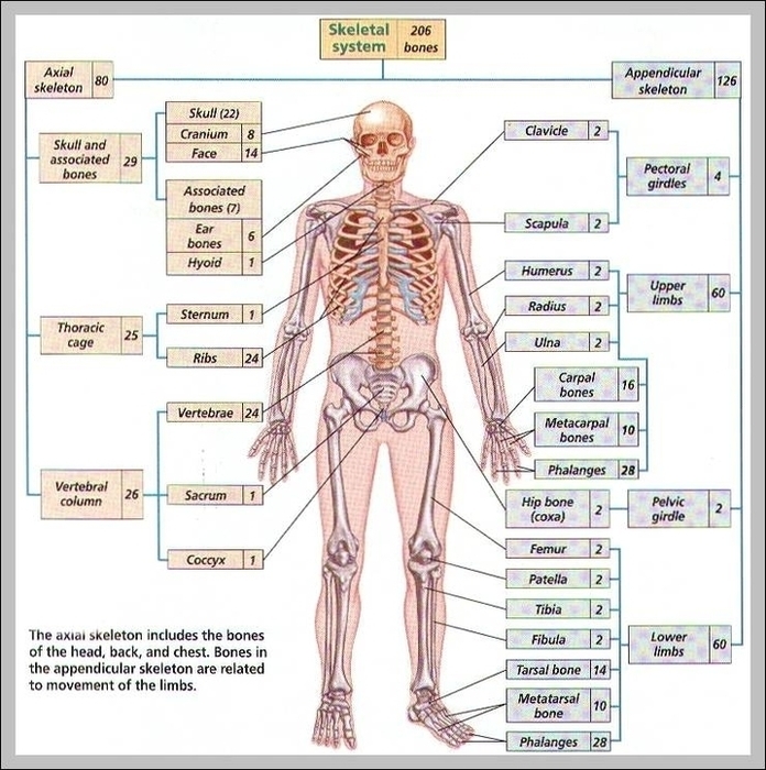
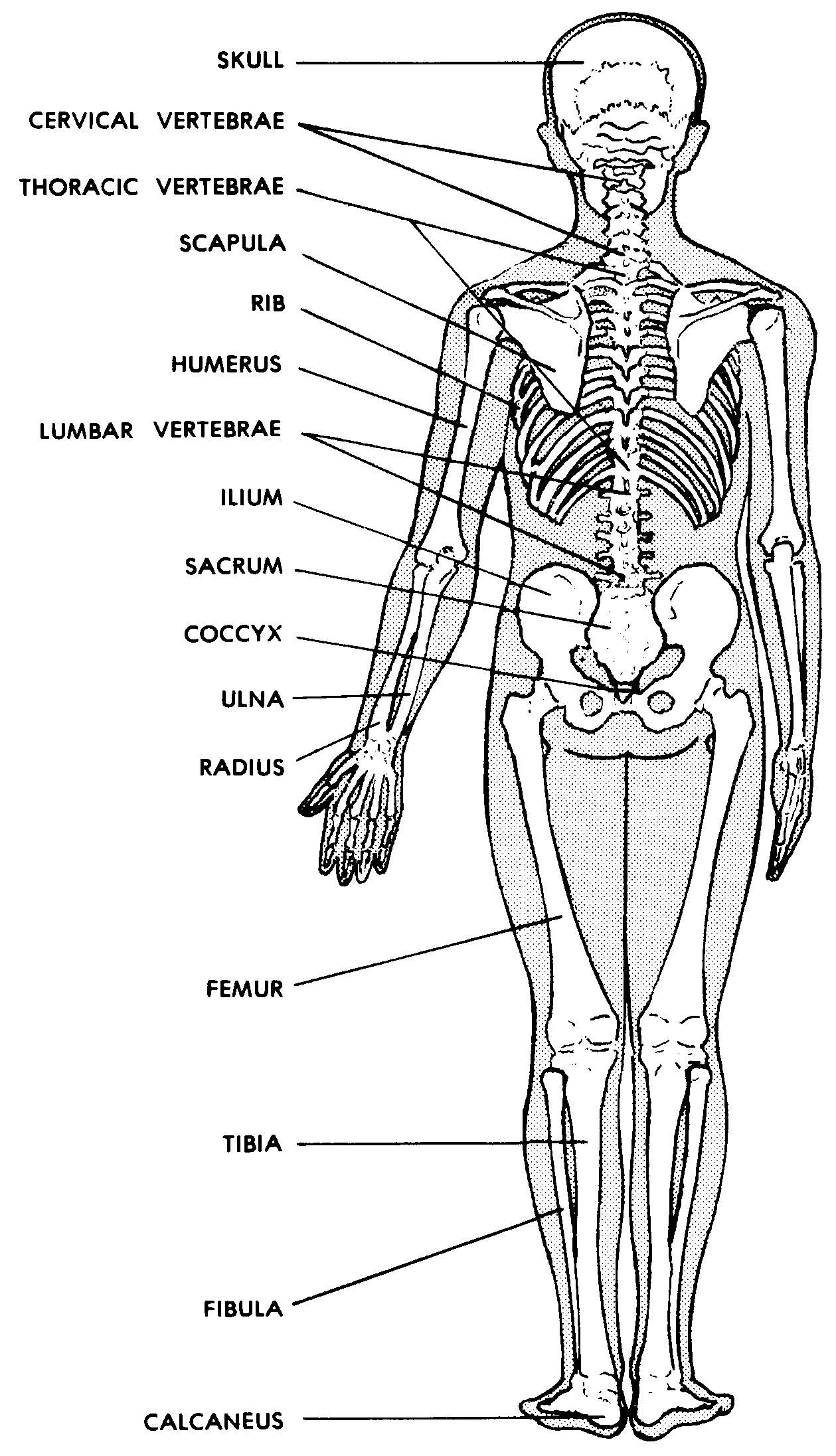
There are 206 bones in the human skeleton, not including teeth and sesamoid bones (small bones found within cartilage): 80 axial bones. This includes the head, facial, hyoid, auditory, trunk, ribs, and sternum. 126 appendicular bones. This includes arms, shoulders, wrists, hands, legs, hips, ankles, and feet.

Major Bones In The Human Body Diagram Major Bones Of The Body Diagram
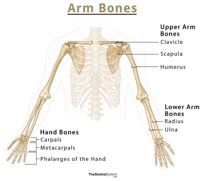
The human skeletal system consists of all of the bones, cartilage, tendons, and ligaments in the body. Altogether, the skeleton makes up about 20 percent of a person's body weight. An adult's.
Human Body Bones Diagram / Human skeleton Hands and feet Britannica
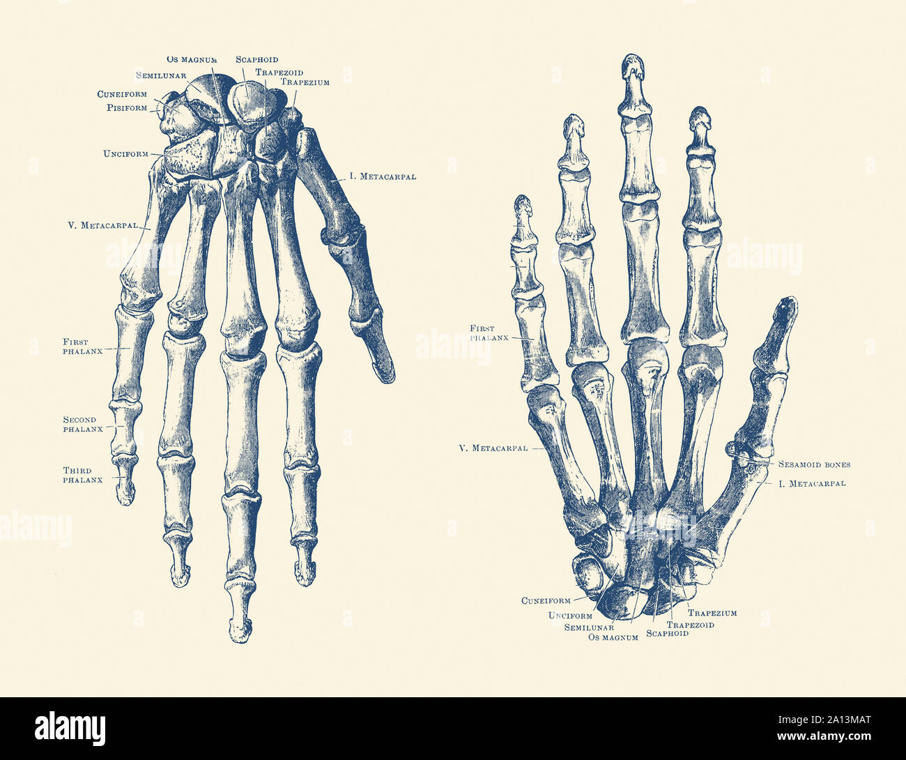
The only short bones in the human skeleton are in the carpals of the wrists and the tarsals of the ankles. Short bones provide stability and support as well as some limited motion. Flat Bones. The term flat bone is somewhat of a misnomer because, although a flat bone is typically thin, it is also often curved. Examples include the cranial.

humanskeletondiagram Tim's Printables
The patella is a triangular bone that forms a protective cap over the knee joint. Also known as the kneecap, it articulates with the femur (thigh bone). It is the largest sesamoid bone in the human body. Tarsal Bones. The tarsal bones are the bones of the ankle, and there are 14 tarsal bones, 7 on each foot. They are as under.

Quick Tips How to Estimate the Chronological Age of a Human Skeleton
human skeleton, the internal skeleton that serves as a framework for the body. This framework consists of many individual bones and cartilages.There also are bands of fibrous connective tissue—the ligaments and the tendons—in intimate relationship with the parts of the skeleton. This article is concerned primarily with the gross structure and the function of the skeleton of the normal.

Printable Human Skeleton Labeled
There are a total of 206 bones in the adult human body. They range in size from the tiniest found in the middle ear, to the largest that forms our thigh.The human body has an amazing array of different bones, many of which you can find on yourself or on a skeleton.Knowledge of the skeletal structure of the human body is essential to know before any anatomy exam, especially in clinical.

34 Human Skeleton With Label Labels For Your Ideas
Definition. Bone is a living, rigid tissue of the human body that makes up the body's skeletal system. Structure. Cortical bone - outer layer. Bone tissue (cancellous bone) - inner layers. Medullary canal - contains either red (active) or yellow (inactive) bone marrow. Types of bones.

Skeletal System Anatomical Chart LAMINATED Human Skeleton Anatomy
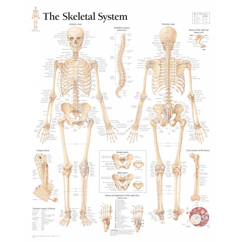
The skeletal system includes over 200 bones, cartilage, and ligaments. Read on to get 10 key facts about the human skeleton. 1. The Skeletal System Consists Of More Than Bones. When you look at the human skeleton the 206 bones and 32 teeth stand out. But look closer and you'll see even more structures.

Human Skull Diagrams 101 Diagrams
Bones are composed of two types of tissue. Compact (cortical) bone is a hard outer layer that is dense, strong, and durable. It makes up around 80% of adult bone mass and forms the outer layer of.
Human Bone Anatomy Chart The Human Skeletal System Anatomical Poster
Bone Structure of the Chest and Hip. The bones shown in the chest and hip region in the labeled human skeleton diagram are the ribs, vertebrae, pelvis, OS coxae, sacrum and coccyx. Total there are 12 pairs of ribs, as you can see in the diagram. The last pair of the ribs, which is at the bottom of the rib, are called floating ribs, as they are.

Human skeleton with labeled bones
Main bones of the skeletal system. We'll begin by looking at the skeletal system. As the name implies, the structural and functional unit is bone-a highly specialized and hard connective tissue. Bones can be classified according to two major criteria, yielding different types of bones:. Compact and spongy bone (according to strength); Long, short, flat, irregular, and sesamoid (according.

206 Bones of the Human Skeleton. But we start with 270 bones! ⋆ Santa
Gross Anatomy of Bones. A long bone has two main regions: the diaphysis and the epiphysis ( Figure 6.3.1). The diaphysis is the hollow, tubular shaft that runs between the proximal and distal ends of the bone. Inside the diaphysis is the medullary cavity, which is filled with yellow bone marrow in an adult.