Alveoli Definition, Location, Anatomy, Function, Diagrams
To "borrow" this diagram from Gill et al (1979), one can see that one's alveoli are only vaguely spherical when they are distended, and in a collapsed state they take on a sorry folded-looking shape. The point of this diagram is to impose upon the reader the significance of lung volume on alveolar shape and surface area changes.

Pulmonary alveolus Wikipedia
Pulmonary alveolus ( plural: alveoli) are tiny air sacs that function as basic respiratory units. It is a hollow cup-shaped cavity in the lung parenchyma, where gas exchange takes place. Lung alveoli are found in the acini at the beginning of the respiratory zone.
:max_bytes(150000):strip_icc()/what-are-alveoli-2249043-01-94dfddd4dfe9488b8056d586824c7c36.png)
Alveoli Function, Structure, and Lung Disorders
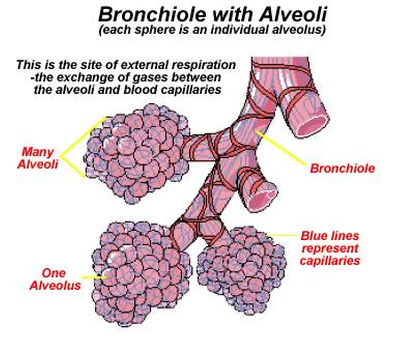
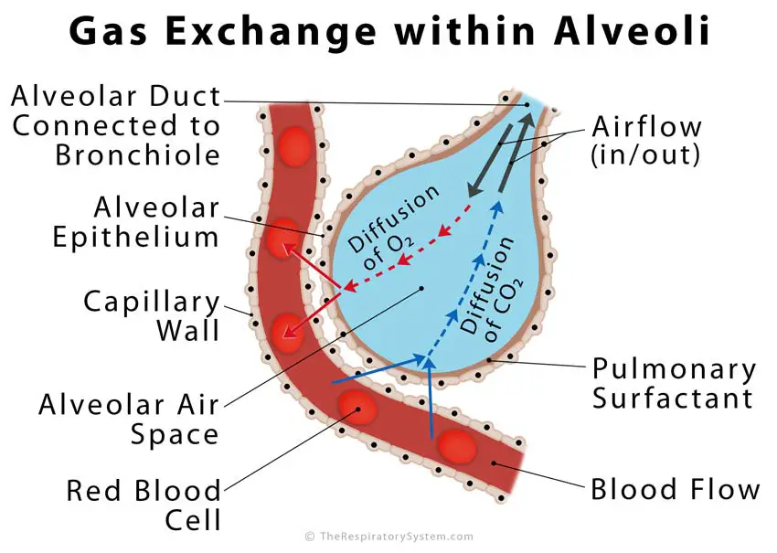
Gas exchange occurs at the alveoli in the lungs and takes place by diffusion. The alveoli are surrounded by capillaries so oxygen and carbon dioxide diffuse between the air in the alveoli and the.
Pictures Of Alveoli
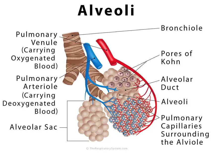
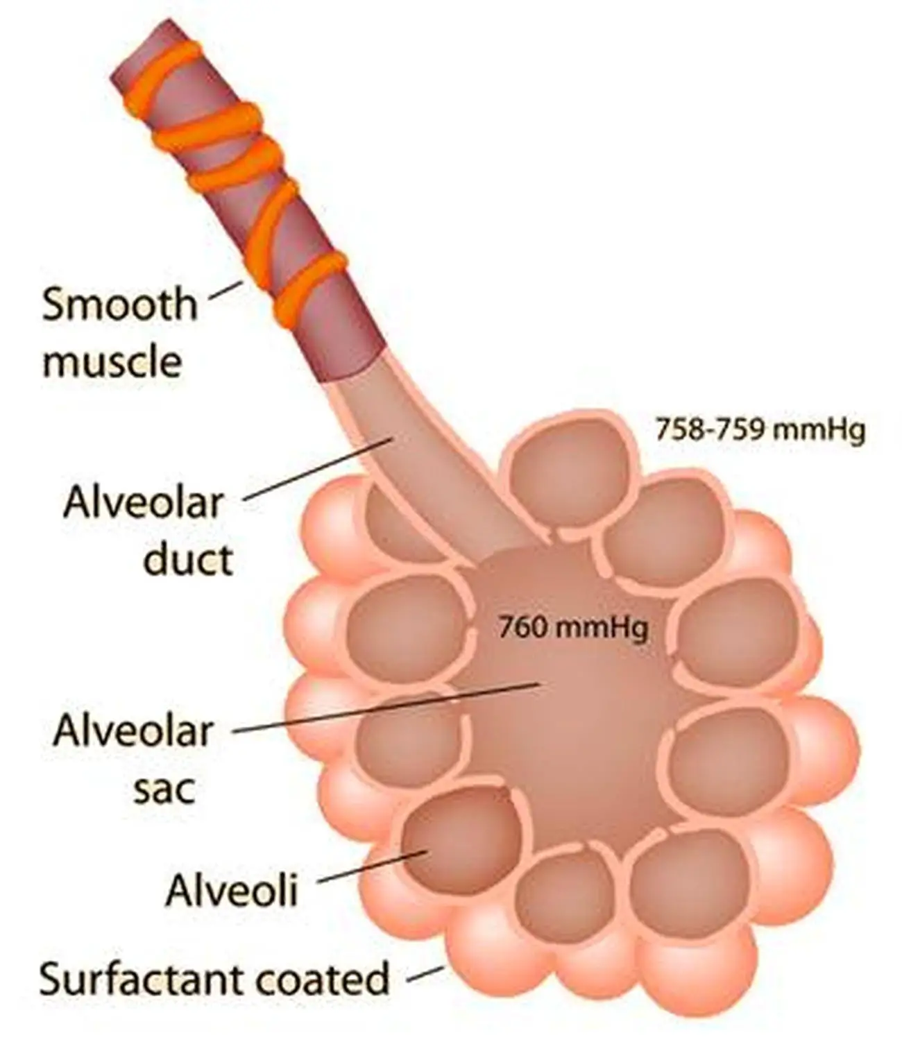
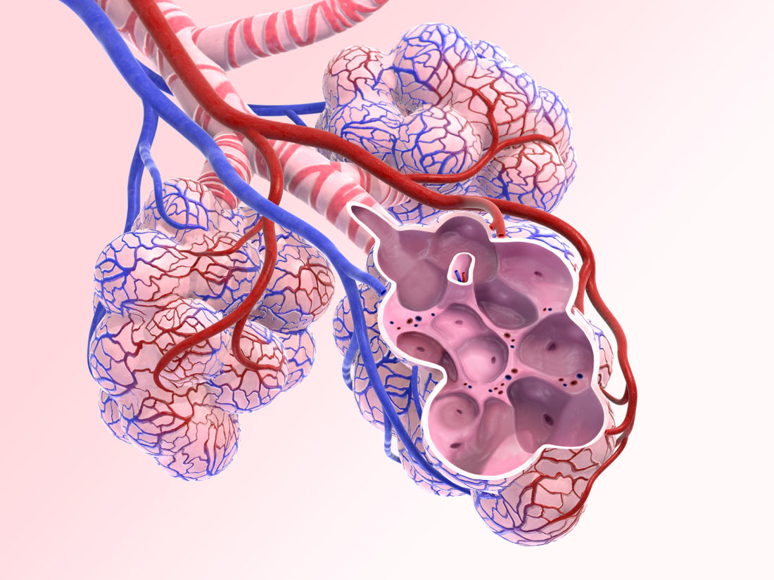
1 2 At the distal end of an alveolar duct, the alveoli are arranged into grape-like clusters called alveolar sac. The alveoli share a common opening to the alveolar duct. Alveolar sacs. Lung Alveolus Structure - Lung Alveoli Anatomy

Alveoli diagram Biology lessons, Nursing mnemonics, Medical surgical
Alveoli are the small balloon-like sacks of 200-500μm diameter [1], making up a vital part of the respiratory zone of the human lungs. Each alveolus (singular) plays an important role in letting oxygen and carbon dioxide move into and from the bloodstream during inhalation and exhalation [2, 3]. Where are the alveoli found
/Alveoli-56a14da83df78cf772696d4f.jpg)
Alveoli Function, Structure, and Lung Disorders
Alveoli represent the major sites of gas exchange. Their presence increases the surface area of the lung to maximize gas exchange, much like villi and microvilli increase the absorptive surface area of the digestive tract. For alveoli to carry out their function efficiently, they must be both a dynamic and stable system. The lung parenchyma must be able to expand and recoil during inspiration.

Respiratory System Anatomy
The term alveolus (singular) refers to a hollow cavity, basin or bowl in latin. Consequently, there are different types of alveoli (plural) found throughout the human body. However, alveoli are most often used to describe the small air sacs of the lungs of mammals, and are therefore known more specifically as the pulmonary alveoli .
Pictures Of Alveoli
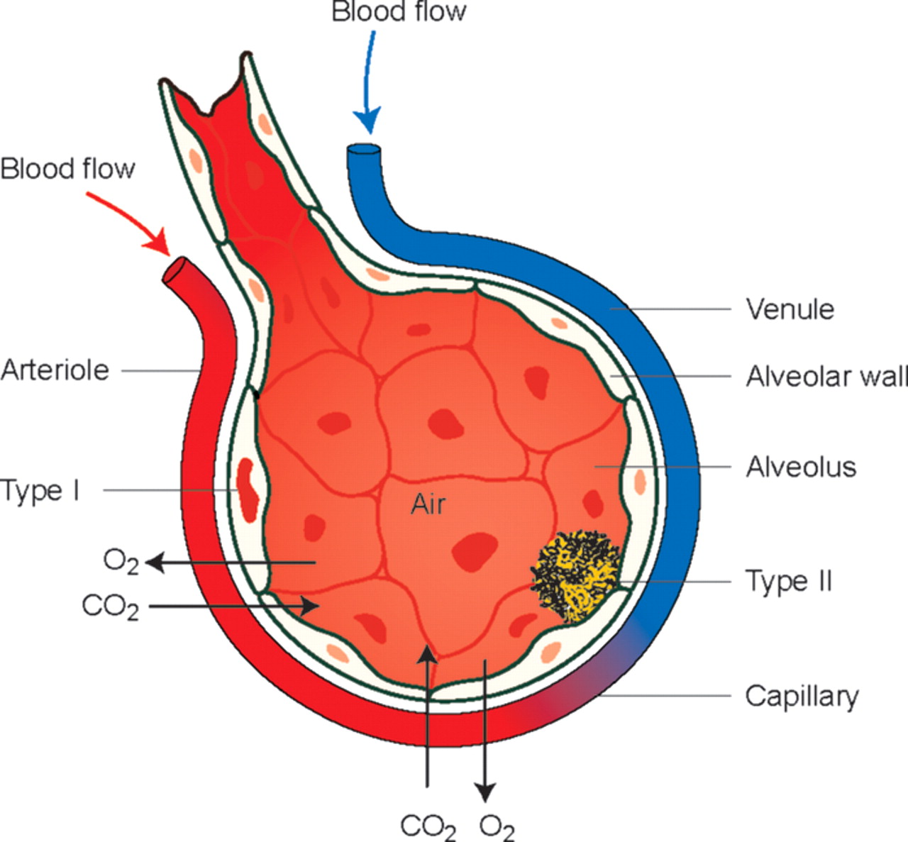
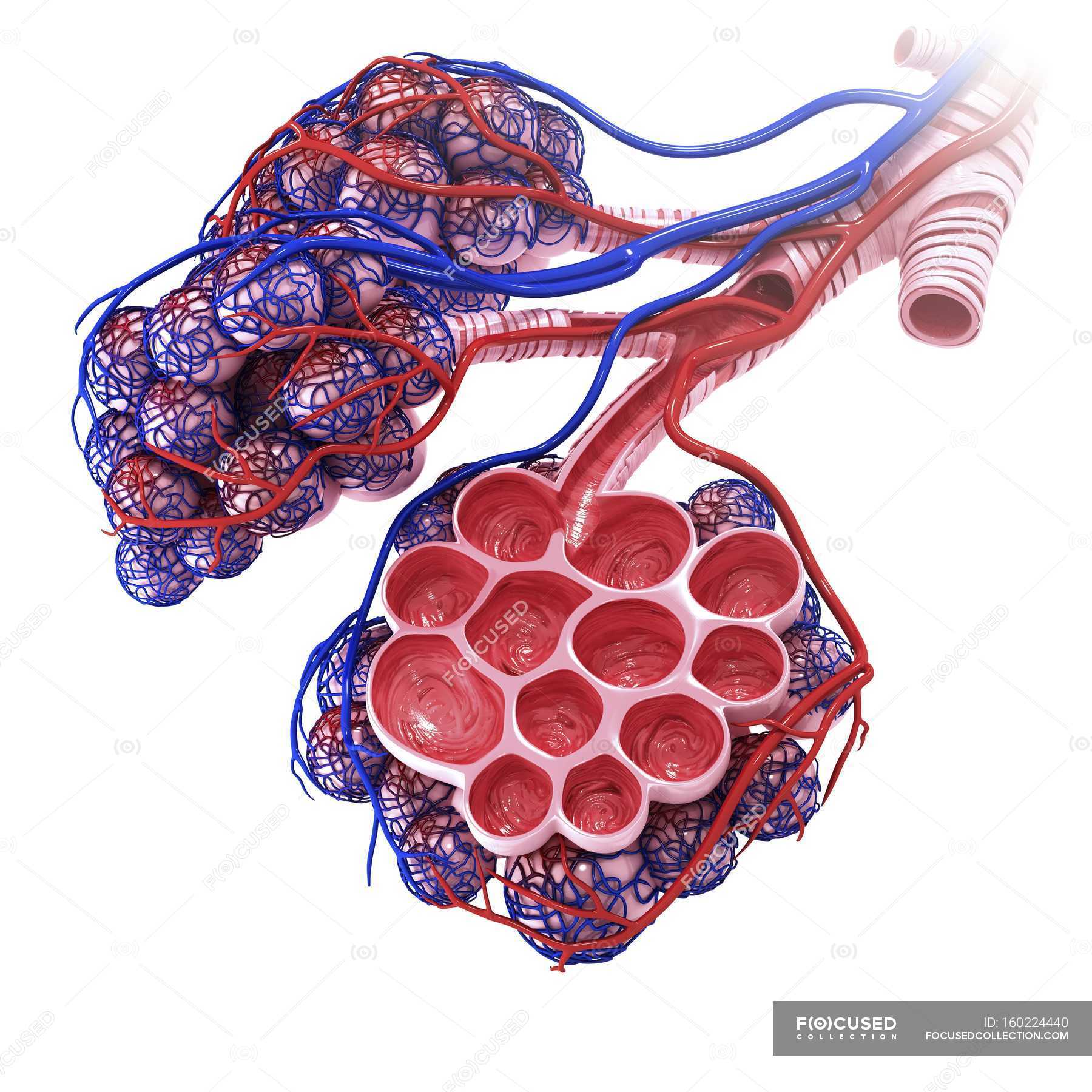
This diagram shows a diagram of an alveolar sac, showing how the organisation of the alveoli, and the network of blood capillaries that surround the alveoli (in red). The epithelium of the alveoli, contains two main types of cells:

Pin on EMT
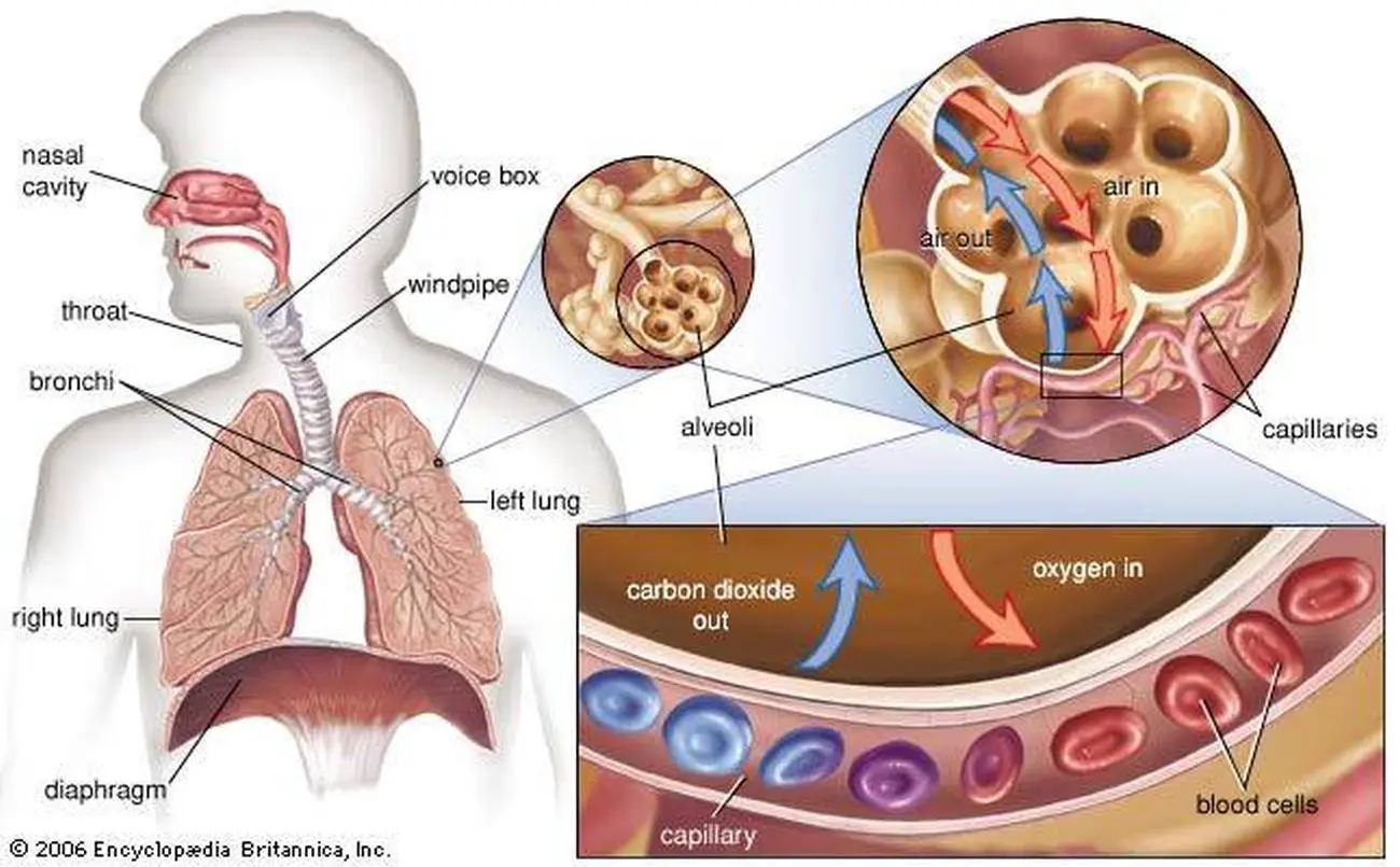
Alveoli are tiny, balloon-shaped air sacs located at the end of the bronchioles, the branch-like tubes in the lungs. The alveoli move oxygen and carbon dioxide (CO 2) molecules into and out of your bloodstream. This article discusses the structure and function of the alveoli.
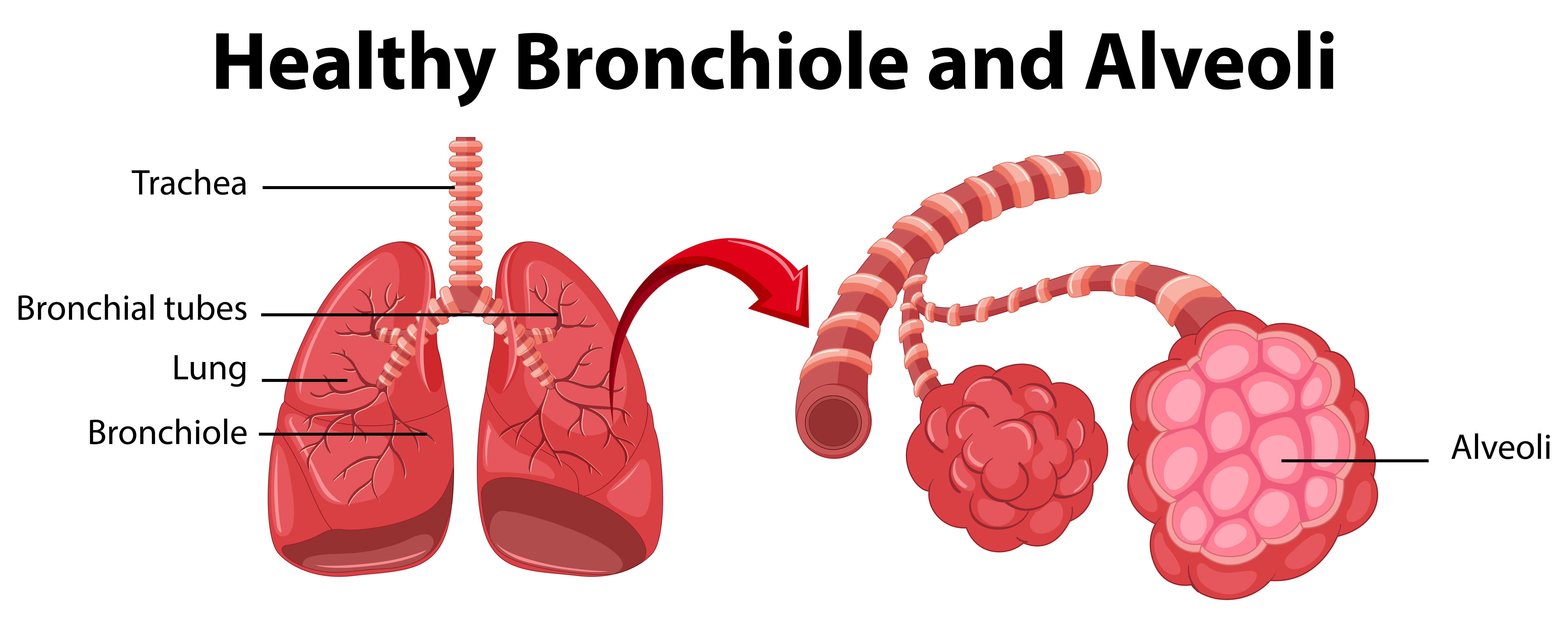
Diagram showing healthy bronchiole and alveoli 434375 Vector Art at
Takeaway Alveoli are tiny air sacs in your lungs that take up the oxygen you breathe in and keep your body going. Although they're microscopic, alveoli are the workhorses of your respiratory.
Alveoli Definition, Location, Anatomy, Function, Diagrams
Large surface area - many alveoli are present in the lungs with a shape that further increases surface area.; Thin walls - alveolar walls are one cell thick providing gases with a short diffusion.
Alveoli crosssection A KYU Design
The alveoli are located in the respiratory zone of the lungs, at the distal termination of the alveolar ducts. These air sacs are at the end points of the respiratory tract. There are approximately 700 million alveoli in the lungs, covering a total surface area of about 70 m 2, which is a considerably larger surface area relative to volume. The.
Pictures Of Alveoli
Alveoli are tiny air sacs in the lungs where gas is exchanged during breathing. Within the human lungs the alveoli provide an efficient exchange surface adapted for gas exchange. This involves the.
Biology Diagrams,Images,Pictures of Human anatomy and physiology
At the end of the bronchioles are air sacs called alveoli,. Diagram labeling the major structures of the respiratory system. Image credit: Arteries and veins of the body by OpenStax, CC BY 4.0. An important structure of respiration is the diaphragm. When the diaphragm contracts, it flattens and the lungs expand, drawing air into the lungs.

Fully Labelled Vector & Photo (Free Trial) Bigstock
The average human adult has around 480 - 500 million alveoli in their lungs. This equals a surface area of 40 - 75 m 2. The large number of alveoli increases the surface area available for oxygen and carbon dioxide to diffuse across. Thin walls. The walls of the alveoli are only one cell thick and these cells are flattened.
Human alveoli anatomy — cut out, plain background Stock Photo
Lower respiratory tract: Composed of the trachea, the lungs, and all segments of the bronchial tree (including the alveoli), the organs of the lower respiratory tract are located inside the chest.