
Scanning Electron Microscope Of Red Blood Cell Foto de stock Getty Images
Red blood cells (RBC) morphologic evaluation through microscopy optical (OM) and SEM, provides information to forecast, evaluate, and monitor the functioning of many organs.. Red blood cells morphology and morphometry in adult, senior, and geriatricians dogs by optical and scanning electron microscopy Front Vet Sci. 2022 Nov 10;9:998438. doi.
Free picture closer look, details, exhibited, red, blood, cells
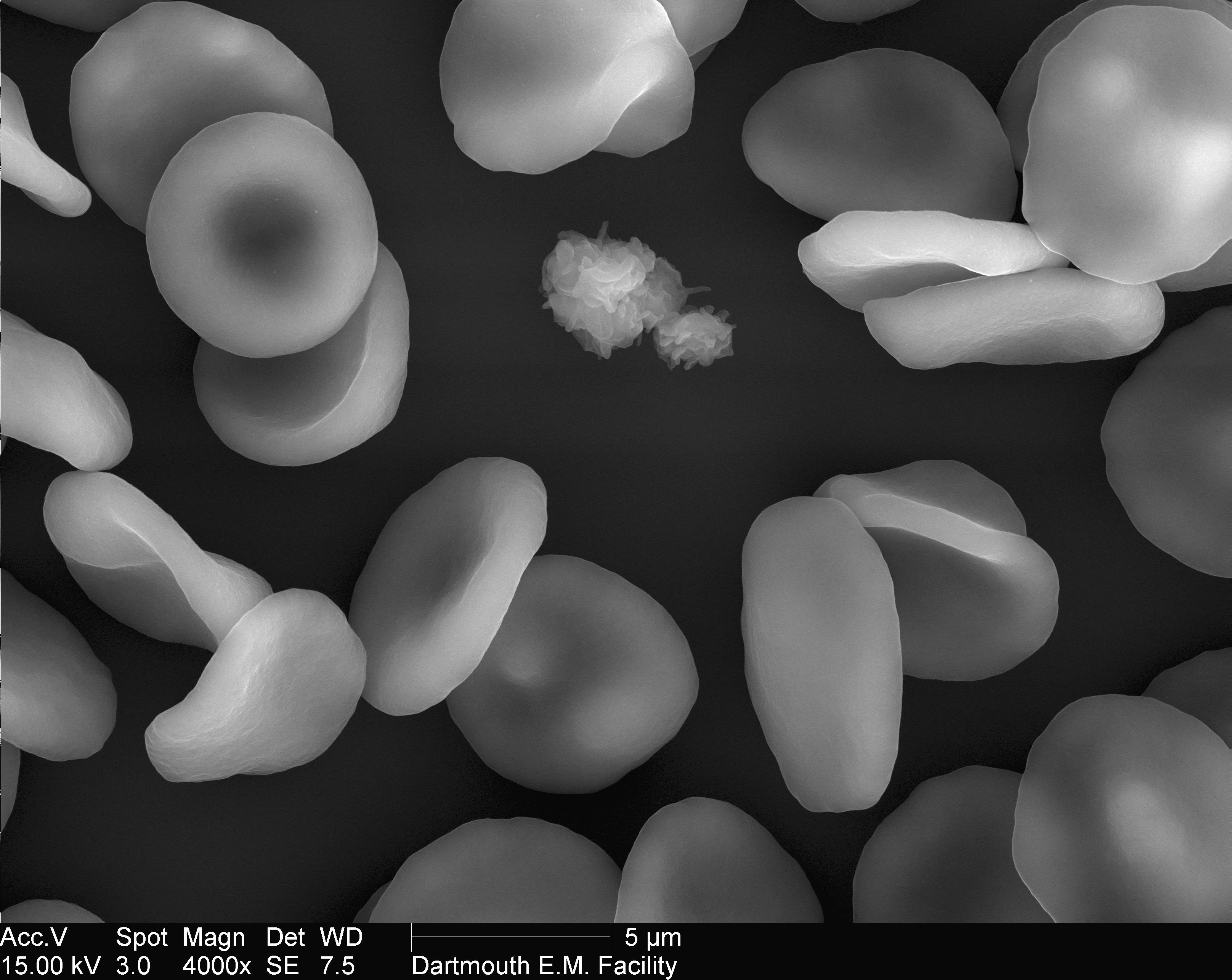
Scanning Electron Microscope Image of Blood Cells: Image Details - NCI Visuals Online Use the search page for more options Use quotation marks (e.g. "breast cancer") to search by that exact term or phrase, no variations. Use multiple keywords separated by spaces (e.g. kidney renal) for broader search results.
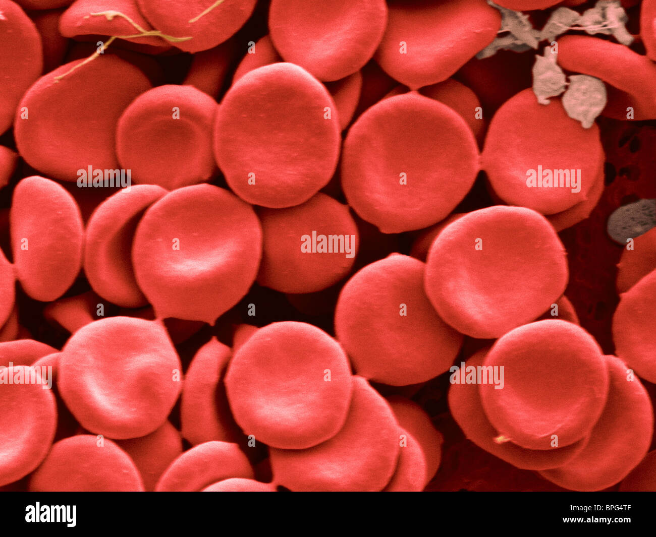
Human red blood cells by scanning electron microscopy (SEM Stock Photo Alamy
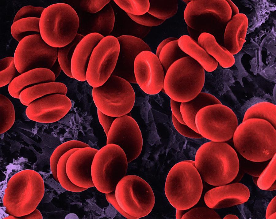
The scanning electron microscope provides a three-dimensional view of surface structure and it has been used for studying human blood cells 1,2. Although this instrument provides much.
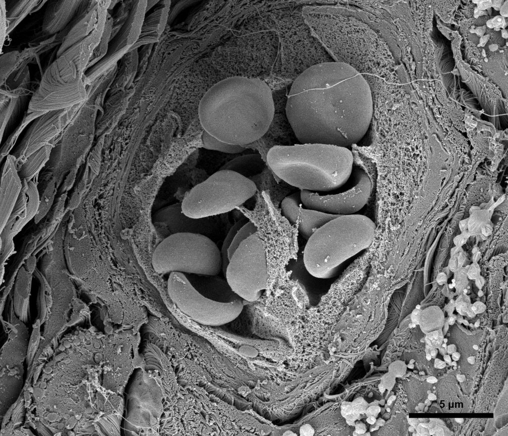
Red Blood Cells in Blood Vessel Bioscience Electron Microscopy Laboratory
The effect of red blood cells (RBC) exposed to an 18 GHz electromagnetic field (EMF) was studied. The results of this study demonstrated for the first time that exposure of RBCs to 18 GHz EMF.
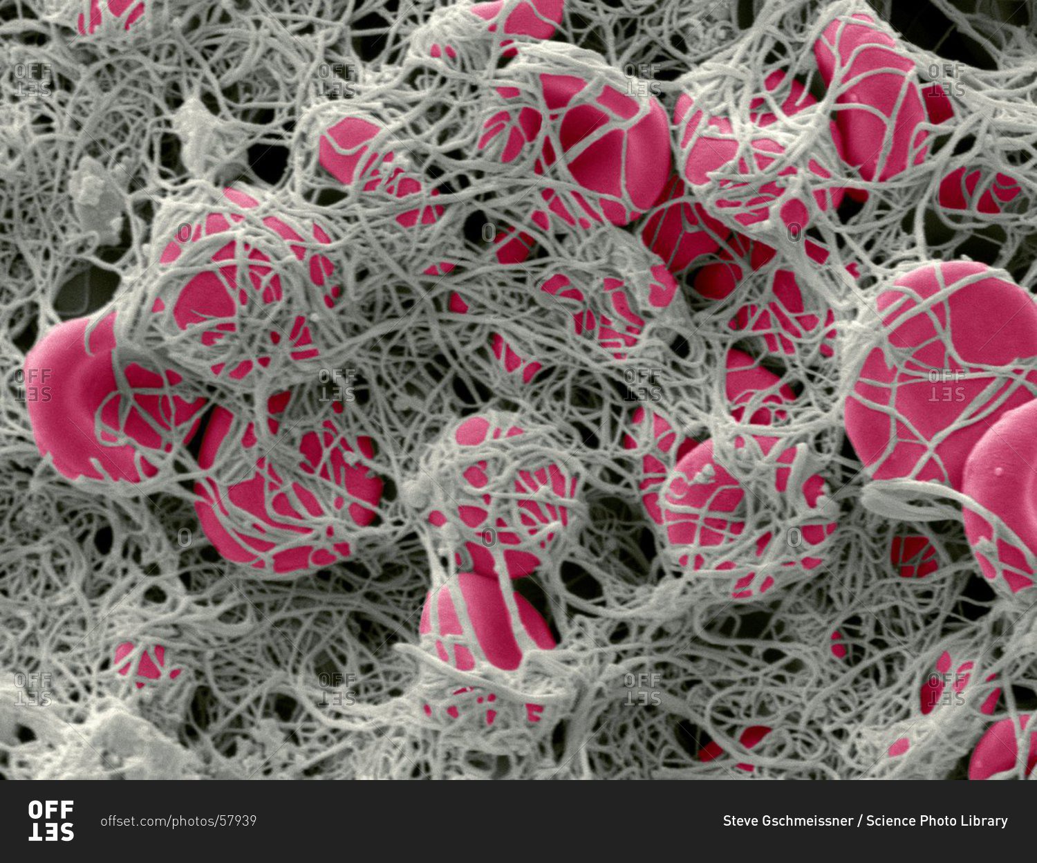
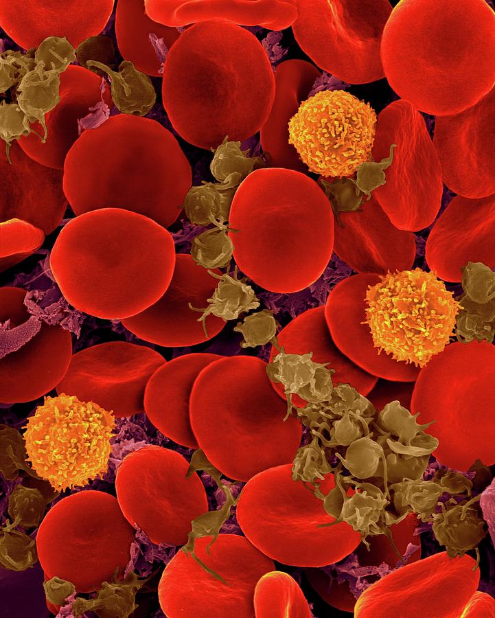
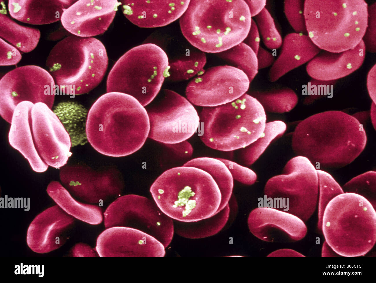
Color scanning electron micrograph of red blood cells (erythrocytes, red) clumped together with
REVIEW published: 01 July 2022 doi: 10.3389/fphys.2022.838071 Light and Scanning Electron Microscopy of Red Blood Cells From Humans and Animal Species Providing Insights into Molecular.

Scanning Electron Microscope Of Red Blood Cell Bildbanksbilder Getty Images
Conventional optical microscopy, scanning electron microscopy (with fixed cells) and transmission electron microscopy (with sections of fixed cells) are extensively used to assess the.
Red blood cells under Electron Microscope u/labduenhas
Visualization of red blood cell morphology by scanning electron microscopy. a Control (without treatment). b Pristine graphene. c Oxidized graphene. d Reduced oxidized graphene. Black arrows point.
Transmission electron micrograph (TEM) of a section of blood vessel full of red blood cells
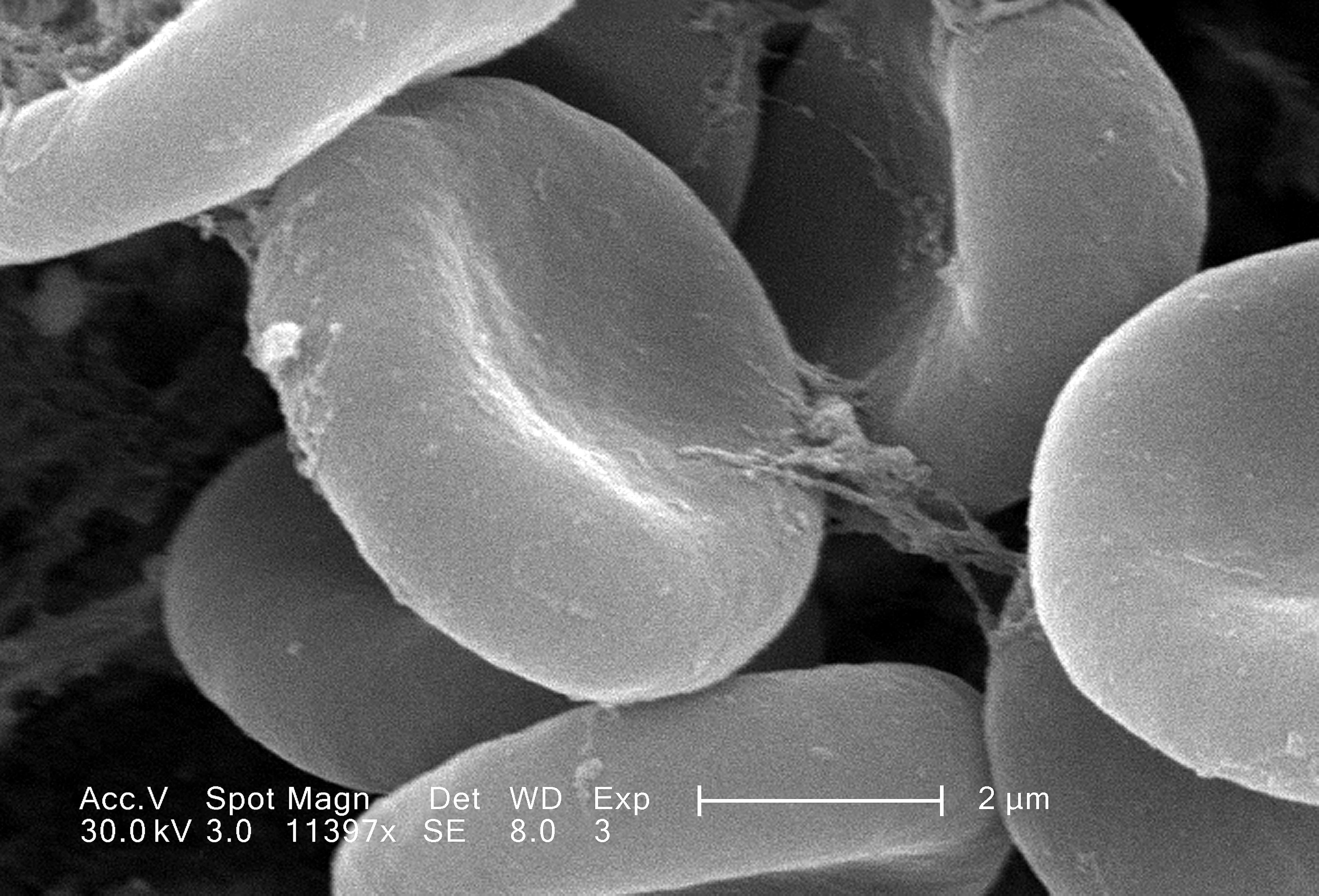
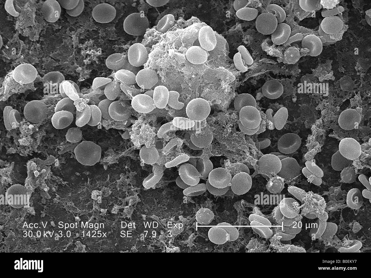
Scanning electron microscopy. A total of 3 ml of whole blood obtained by jugular venipuncture was collected and deposited in tubes with K2EDTA BD Vacutainer ®, which contains the optimal amount of di-potassium EDTA applied by spray to anticoagulant the specified volume of blood (BD Franklin Lakes NJ USA).Once obtained the blood sample was centrifuged at 2,000 rpm for 10 min to separate the.
Red Blood Cells Photograph by Dennis Kunkel Microscopy/science Photo Library Pixels
The ultrastructural anatomy of the blood cells is helpful in the diagnosis of several diseases by the examination of blood cells' morphology at higher magnification using electron microscopes. Diagrammatic representation of various blood cells in the humans and animals.

The erythrocytes, SEM (With images) Scanning electron microscopy, Scanning electron micrograph
Light and Scanning Electron Microscopy of Red Blood Cells From Humans and Animal Species Providing Insights into Molecular Cell Biology CC BY 4.0 Authors: Gheorghe Benga Guy Cox.
Leukemia blood cells under a Color scanning electron micrograph. Red blood cells (erythorocytes
Erythrocytes, red blood cells (RBC), are the functional component of blood responsible for the transportation of gases and nutrients throughout the human body.. Schnitzer B, Rucknagel DL, Spencer HH, Aikawa M. Erythrocytes: pits and vacuoles as seen with transmission and scanning electron microscopy. Science. 1971 Jul 16; 173 (3993):251-2.

(223) Twitter Microscopic photography, Scanning electron microscopy, Scanning electron micrograph
Thanks to Biology Araceli Adabache Ortíz, head of the Scanning Electron Microscopy Laboratory for her invaluable help in obtaining micrographs of red blood cells by SEM, and thanks also to AQB. Sonia Sofía Cruz Muñoz from the Laboratory of Histology and Embryology of the Autonomous University of Aguascalientes for her support in the staining and preservation techniques of blood smears.
Fri., Jan. 18 notes
2 PMID: 35845990 PMC9283769 10.3389/fphys.2022.838071 We reviewed the many discoveries in cell biology, made since the 17 century, which have been based on red blood cells (RBCs).
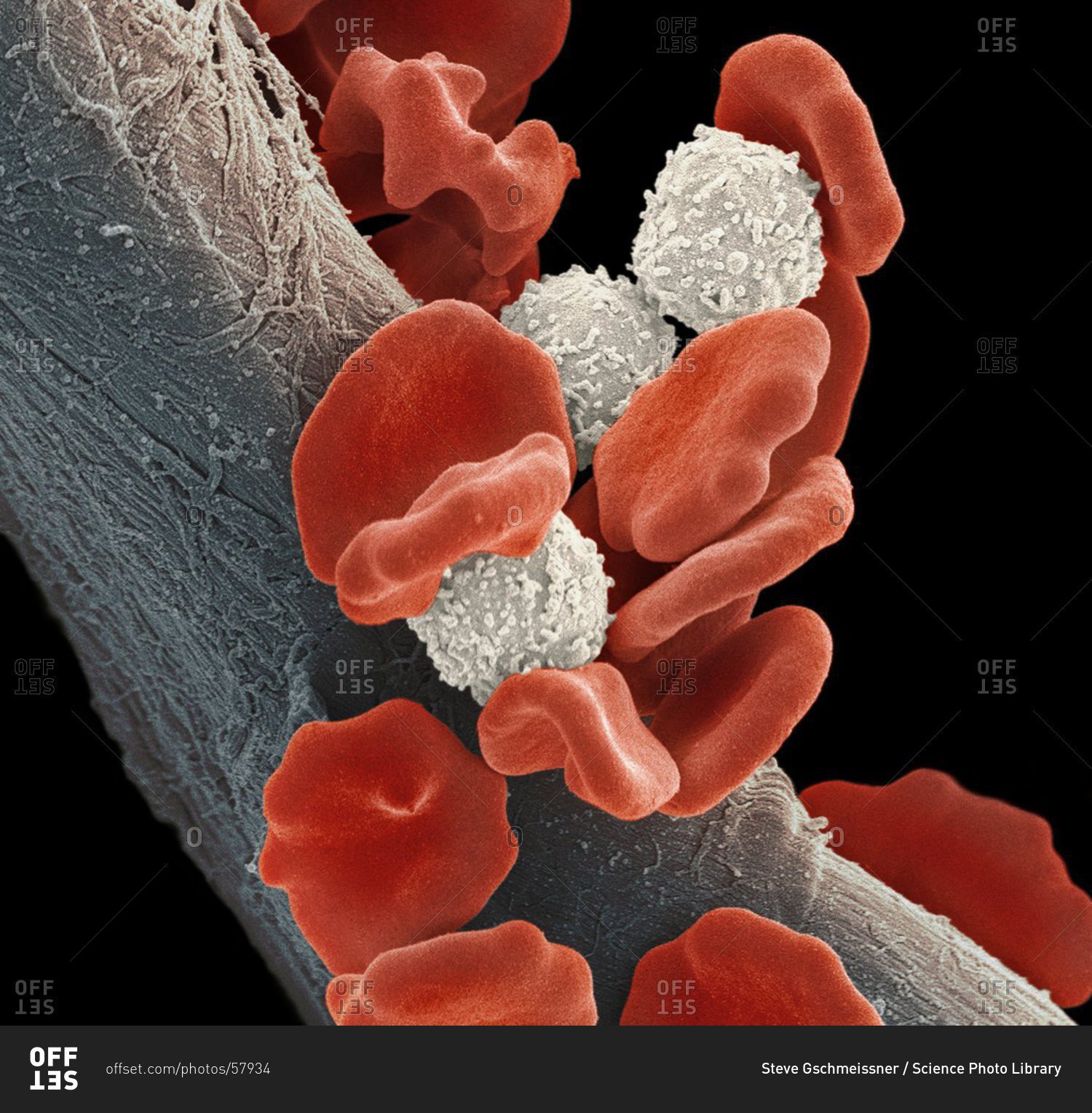
scanning electron microscope image of a blood clot showing red cells and fibrin coagulum Stock
Samples of red of blood cells (RBC), washed free of plasma, from eleven marsupial species were examined in a Jeol JSM-6300 F scanning electron microscope. The diameters of the RBC, lying completely flat or exactly on edge, were measured on photographs using a binocular enlarging optical system with a calibrated eye piece. RBC from the following species were studied: bandicoot (Isoodon.
Red Blood Cells In Isotonic Solution Photograph by Dennis Kunkel Microscopy/science Photo
The Dutch microscopist, Antoni van Leeuwenhoek (1632-1723), is credited by many (e.g., De Robertis, 1970) with this discovery. In a critical analysis of the discovery of blood cells, Hajdu, (2003) concluded that the Dutch naturalist Jan Swammerdam (1637-1680) was the first person to observe RBCs under the microscope.
Red Blood Cells Scanning electron microscope Stock Photo Alamy
PMCID: PMC8914789 PMID: 35271203 Investigation of Red Blood Cells by Atomic Force Microscopy Viktoria Sergunova, 1,* Stanislav Leesment, 2 Aleksandr Kozlov, 3 Vladimir Inozemtsev, 1 Polina Platitsina, 4 Snezhanna Lyapunova, 1 Alexander Onufrievich, 5 Vyacheslav Polyakov, 2 and Ekaterina Sherstyukova 1,3 Bruno Tiribilli, Academic Editor