
Draw the L.S of kidney and label the parts.
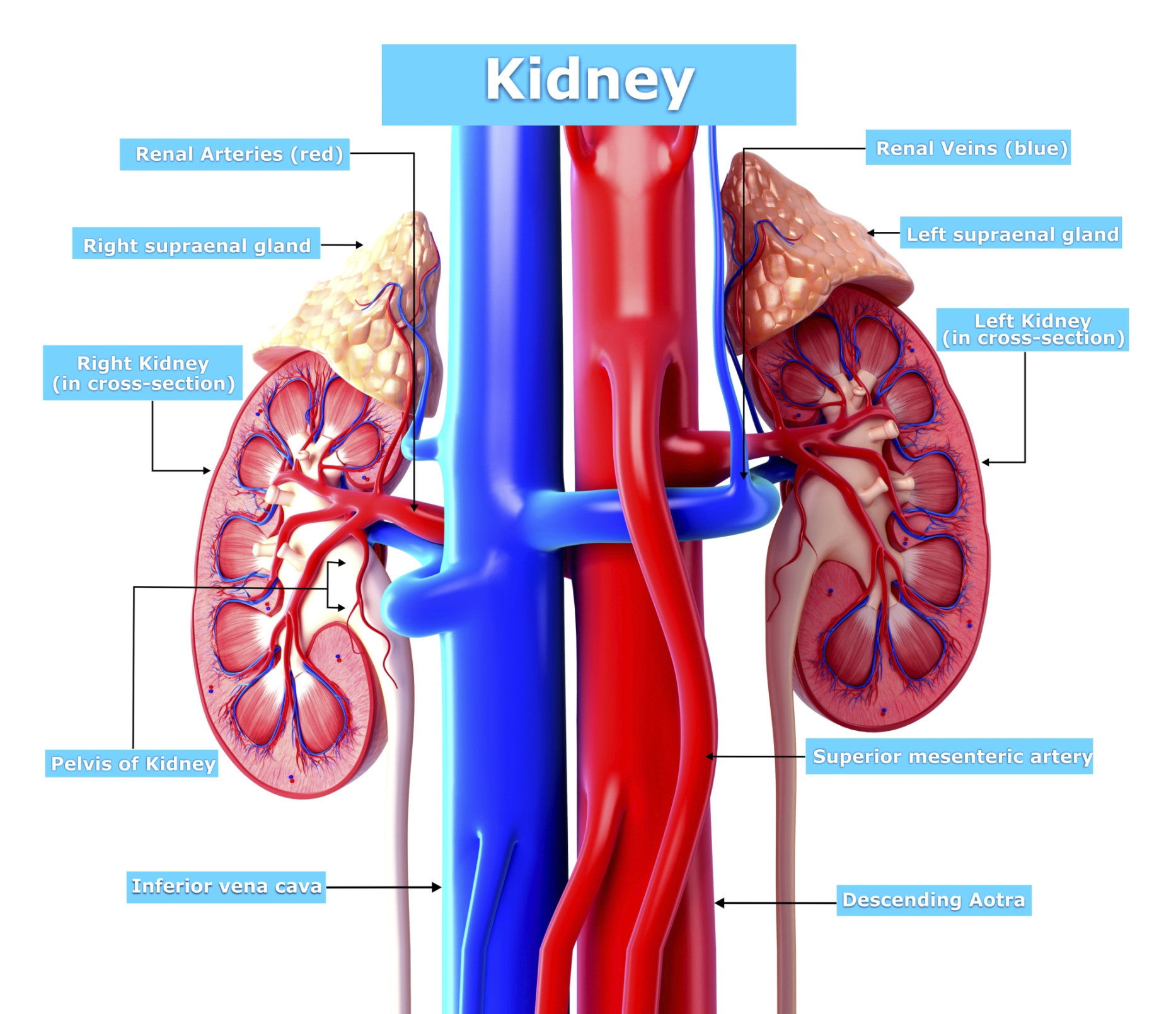
Anatomy Each person has two kidneys. The kidneys are located on either side of the spine, with the top of each kidney beginning around the 11th or 12th rib space. The kidneys are sandwiched between the diaphragm and the intestines, closer to the back side of the abdomen.

Kidney Structures Learn Surgery Online
That this, it is the kidney nephrons that actually perform the kidney's main functions. There are approx. a million nephrons within each kidney. To find out more about these, visit the page about kidney nephrons. Collecting Duct (Kidney): The collecting duct labelled in the diagram above is part of the kidney nephron (shown much enlarged).

Please send me a diagram of L.S Of kidney and label the main parts of it.. Brainly.in
Kidney diagram Kidney conditions Warning signs Health tips What are kidneys? The kidneys are two bean-shaped organs in the renal system. They help the body pass waste as urine. They also.
5b2 Organs HumanBio
Nephrons - Diagram and Structure Nephron is a microscopic structure that is the functional unit of the kidney. There are about 1,000,000 nephrons in each human kidney. Let us look at the components of the nephron. Below is a well-labelled and easy diagram of the nephron for your better understanding.
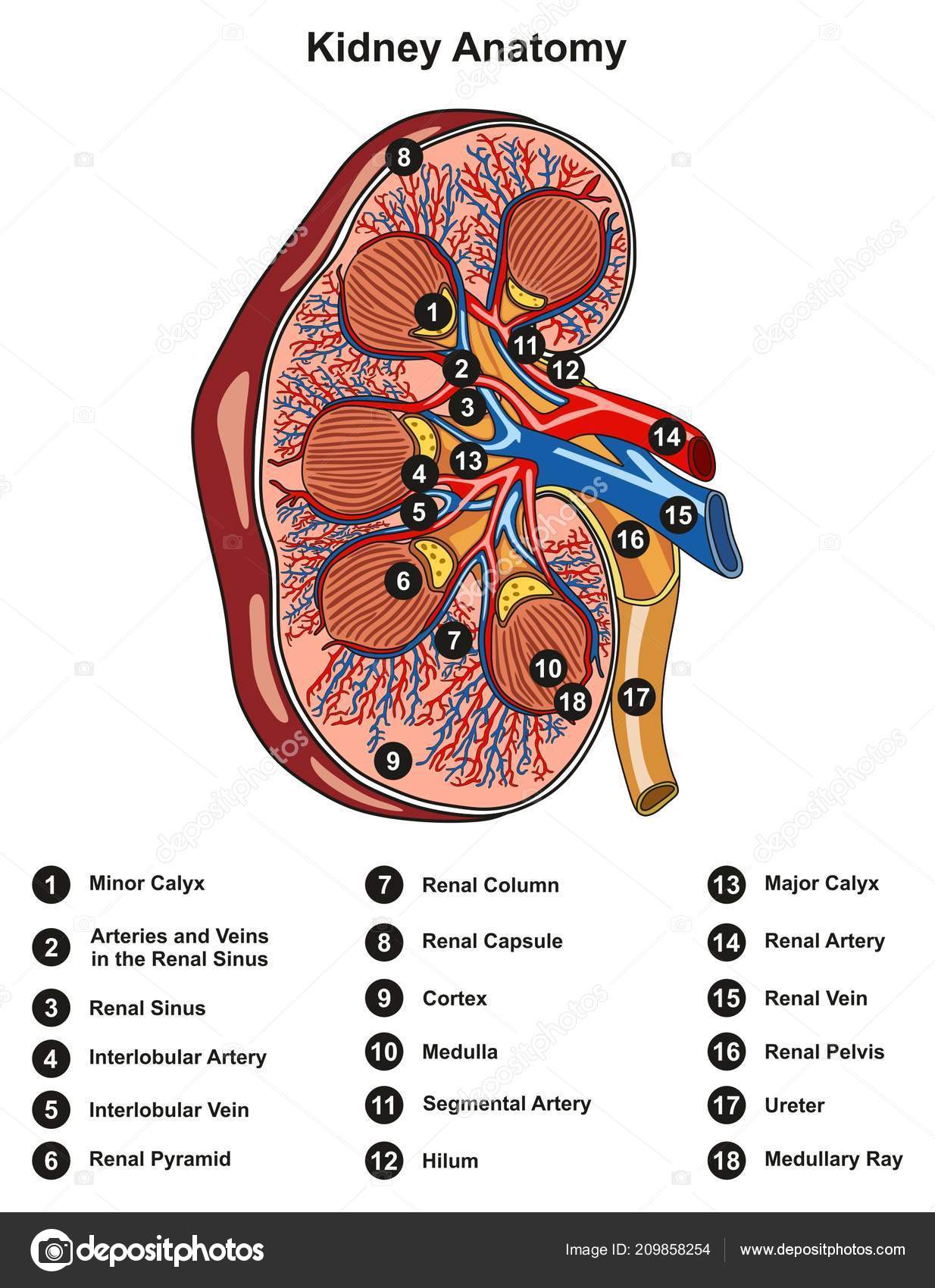
Labeled Kidney Anatomy Cross Section Infographic Diagram Including All Parts Stock Illustration
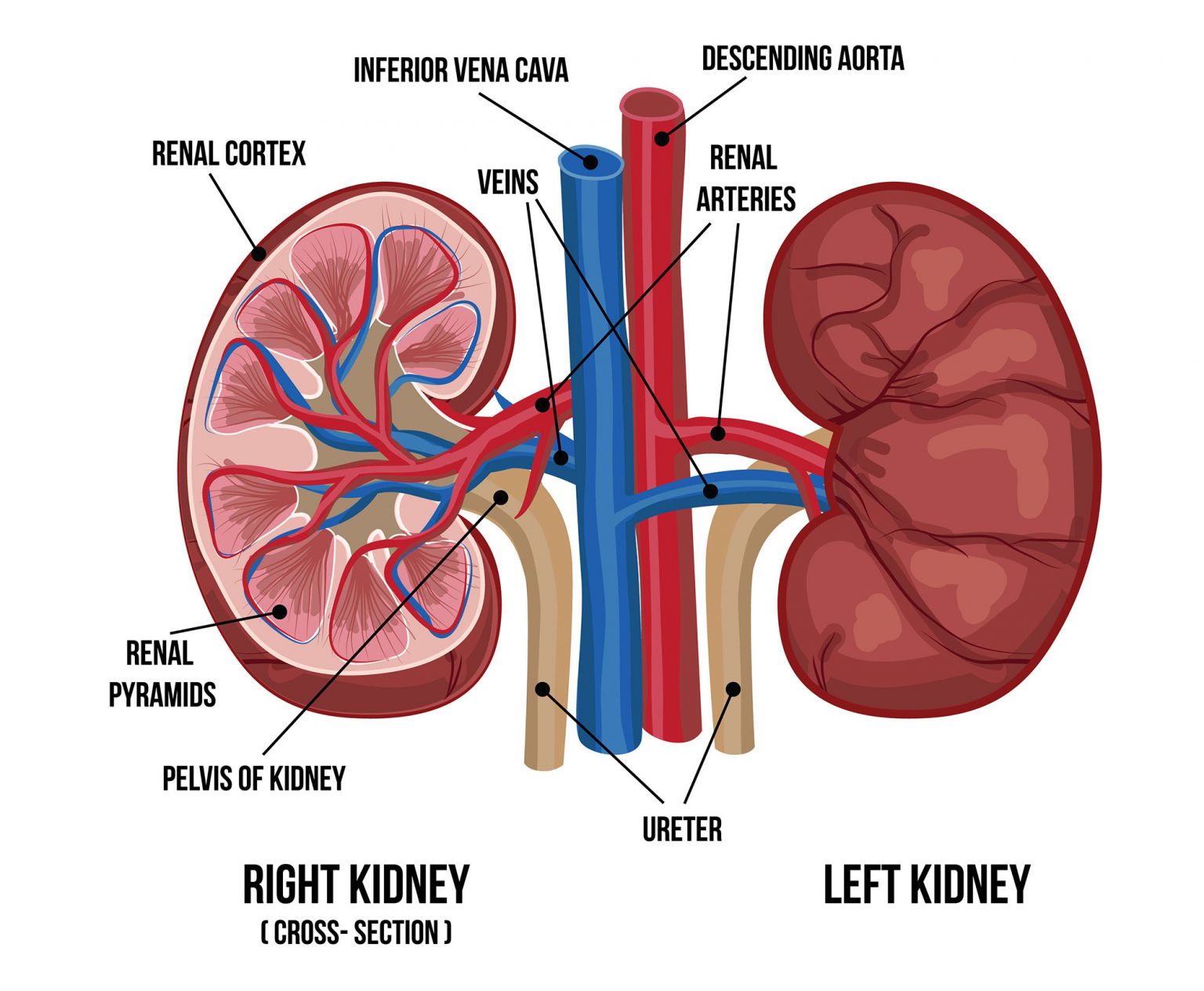
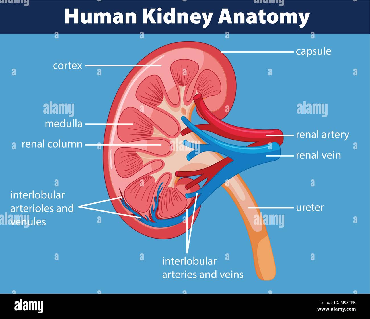
Human kidney is like that of a goat or sheep. If a longitudinal section of the kidney is made by cutting with a long knife from the outer convex surface to the hilum three layers are seen. (1) Outer Cortex (2) Medulla (3) Pelvis 1. Outer Cortex: This is the solid part of the kidney which is dark coloured and granular cortical portion. 2. Medulla:

Cross Section of Right Kidney Stock Image F031/6574 Science Photo Library
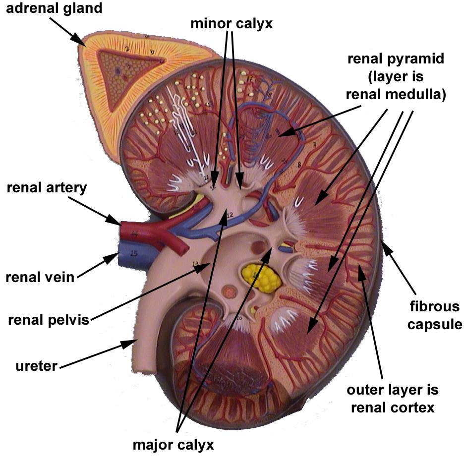
Each kidney weighs about 125-175 g in males and 115-155 g in females. They are about 11-14 cm in length, 6 cm wide, and 4 cm thick, and are directly covered by a fibrous capsule composed of dense, irregular connective tissue that helps to hold their shape and protect them.
Human kidney anatomy diagram 434204 Vector Art at Vecteezy
The video provides a detailed overview of the kidney's smallest functional unit, the nephron. It explains how the nephron filters blood, excretes waste, and maintains water levels. The video also highlights the role of the renal cortex and renal medulla in this process. The loop of Henle's function in reabsorbing water and making the renal.
Human Kidney Cross Section, Scientific Background, Anatomy, Urinary System with Main Parts
Each kidney weighs about 125-175 g in males and 115-155 g in females. They are about 11-14 cm in length, 6 cm wide, and 4 cm thick, and are directly covered by a fibrous capsule composed of dense, irregular connective tissue that helps to hold their shape and protect them.

Kidney Anatomy infographic LifeMap Discovery
nephron, functional unit of the kidney, the structure that actually produces urine in the process of removing waste and excess substances from the blood.There are about 1,000,000 nephrons in each human kidney. The most primitive nephrons are found in the kidneys of primitive fish, amphibian larvae, and embryos of more advanced vertebrates.The nephrons found in the kidneys (mesonephros) of.
Human Kidney Medical Diagram High Resolution Stock Photography and Images Alamy
Each nephron consists of a blood supply and a specialized network of ducts called a tubule. For each nephron, an afferent arteriole feeds a high-pressure capillary bed called the glomerulus. Blood is filtered by the glomerulus to produce a fluid which is caught by the nephron tubule, called filtrate. The proximal end of the tubule that.

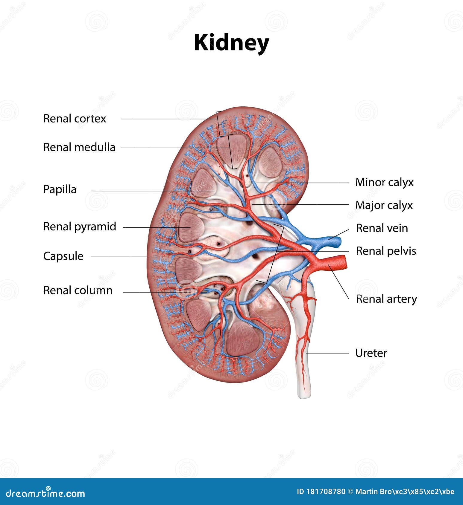
Normal Kidney Anatomy
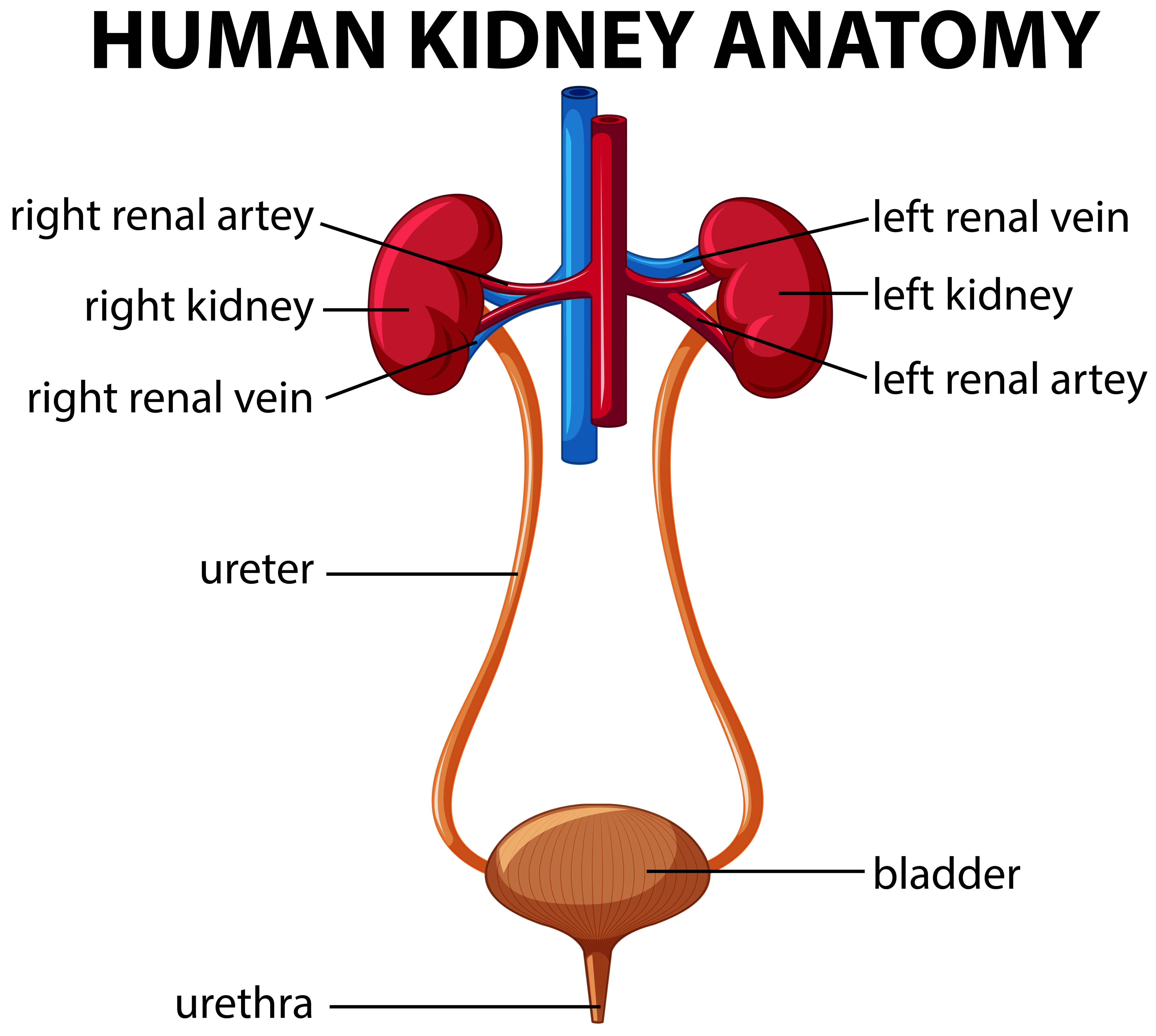
The system contains two kidneys, which control the electrolyte composition of the blood and eliminate dissolved waste products and excess amounts of other substances from the blood; the latter substances are excreted in the urine, which passes from the kidneys to the bladder by way of two thin muscular tubes called the ureters.

Kidney diagram simple Healthiack
Structure. Each of the two bean-shaped organs weighs about 125 to 175 grams and 115 to 155 grams in males and females respectively. The kidney typically measures approximately 11 to 14 centimeters.
Human kidney anatomy diagram 446409 Vector Art at Vecteezy
In humans, the kidneys are two reddish-brown bean-shaped blood-filtering organs [1] that are a multilobar, multipapillary form of mammalian kidneys, usually without signs of external lobulation. [2] [3] They are located on the left and right in the retroperitoneal space, and in adult humans are about 12 centimetres ( 41⁄2 inches) in length.
Biology (MBBS) Structure of Human Kidney with labeled diagram Ratta.pk
Reading time: 23 minutes Recommended video: External structure of the kidney [11:47] Overview of the structure of the kidney. Kidney Ren 1/3 Synonyms: none The kidneys are bilateral organs placed retroperitoneally in the upper left and right abdominal quadrants and are part of the urinary system.

Human kidney anatomy. External view (a), internal view (b), and its... Download Scientific Diagram
Anatomical Position. The kidneys lie retroperitoneally (behind the peritoneum) in the abdomen, either side of the vertebral column.. They typically extend from T12 to L3, although the right kidney is often situated slightly lower due to the presence of the liver.Each kidney is approximately three vertebrae in length. The adrenal glands sit immediately superior to the kidneys within a separate.
What Are the Parts of the Human Kidney? Healthfully
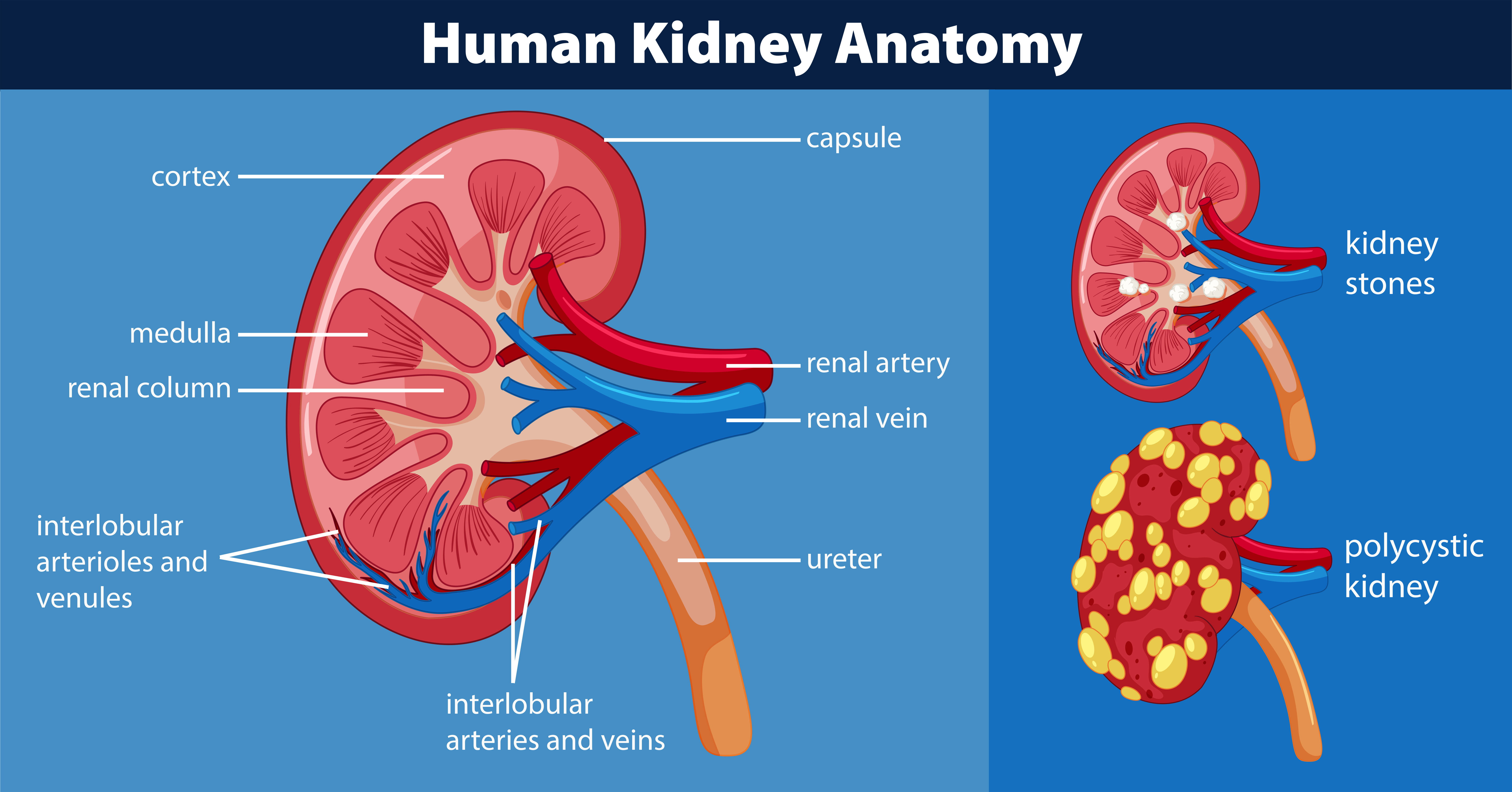
A simple structure of kidney can be understood by the following diagram: Function The most important function of the kidney is to filter the blood for urine formation. It excretes metabolic wastes like urea and uric into the urine. It secretes a number of hormones and enzymes such as: Erythropoietin: It is released in response to hypoxia