
Simple Diagram Of Human Eye With Labelling Human Eye Diagram Class 10 How To Draw Human Eye
1 The human Eye 1.1 Construction of the Eye 1.2 Working of the Eye 1.3 Function of Iris and Pupil 2 Rods and Cones 3 Blind Spot 4 Persistence of Vision 5 Range of Vision of a Normal Human Eye 6 Defects of the Eye 7 Care of the Eyes 8 Visually Challenged Persons Can Read and Write The human Eye

OUR EYES WORK LIKE CAMERA’S! Discovery Eye Foundation
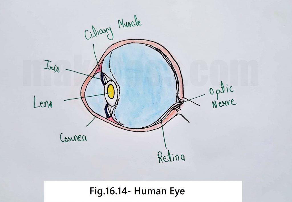
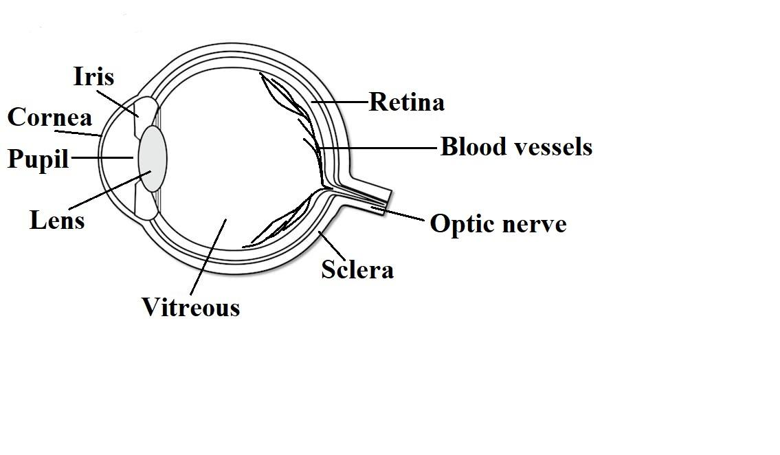
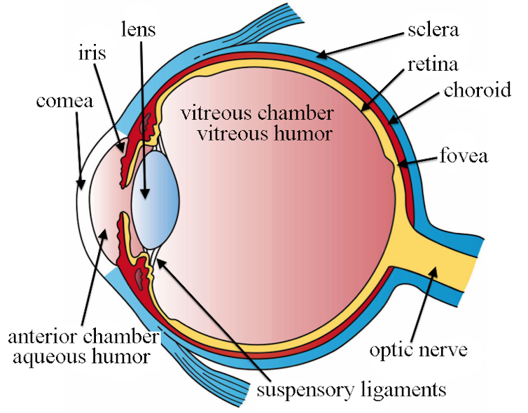
The human eye is a large spherical ball which consists of Crystalline lens Aqueous Humour Pupil Iris Cornea Ciliary Muscles Retina Optic Nerve Vitreous Humour Next: NCERT Question 11 Important → Ask a doubt Class 8 Chapter 16 Class 8 - Light Tired of ads? Get Ad-free version of Teachoo for ₹ 999 ₹499 per month NCERT Questions
Science Class 8 Chapter 16 Light NCERT exercise solution
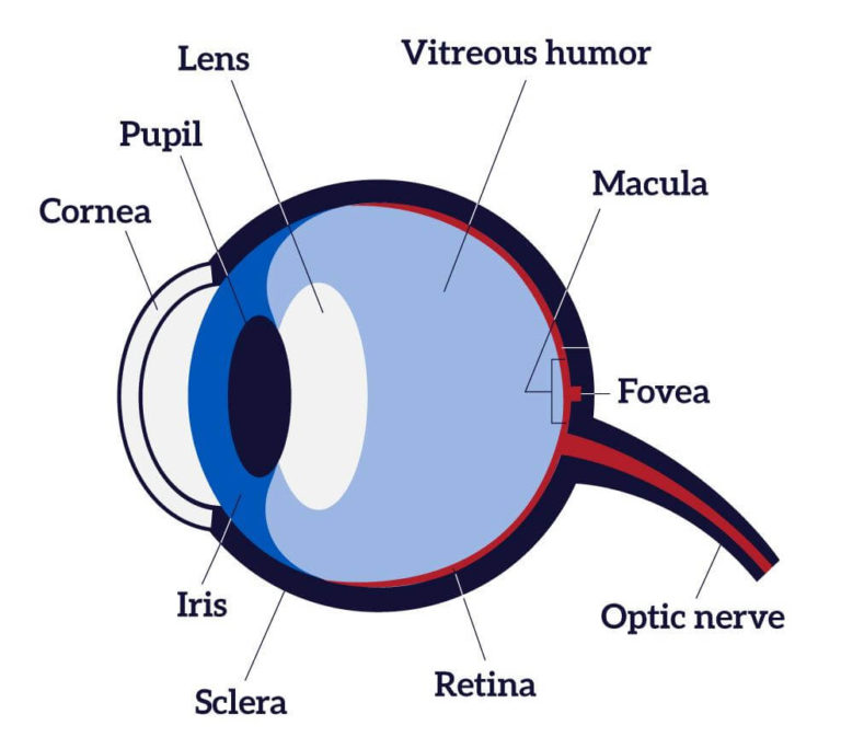
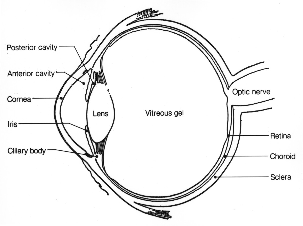
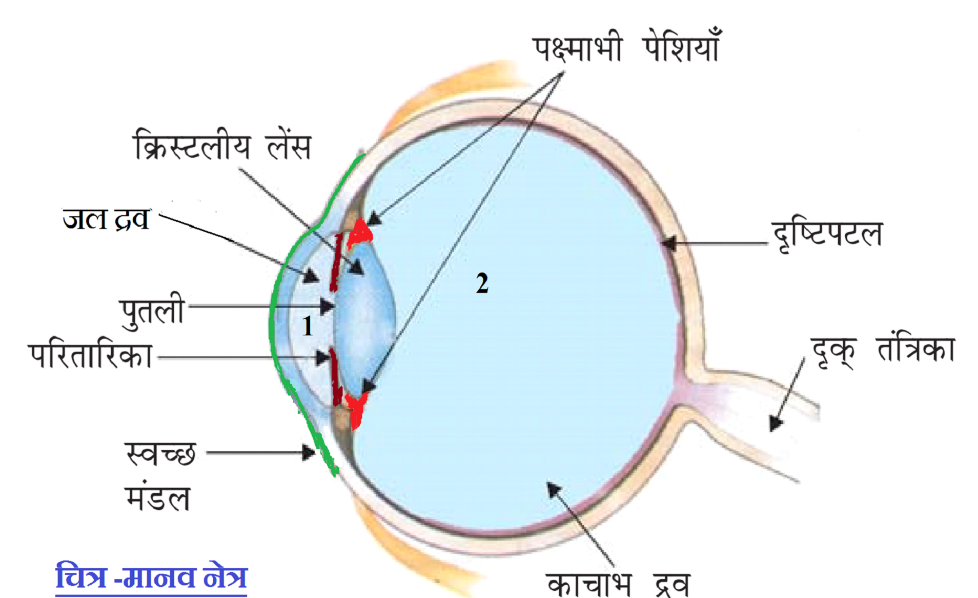
A simplified anatomy of the eye is shown in the diagram below. Parts of Eye Cornea : It is the transparent spherical membrane covering the front of the eye. Iris : It is the coloured diaphragm between the cornea and lens. Pupil : It is the small hole in the iris. Eye lens : It is a transparent lens made of jelly like material.

Anatomy of the Eye Editable PowerPoint Presentation/Main parts of Human Eye /Free PPT YouTube
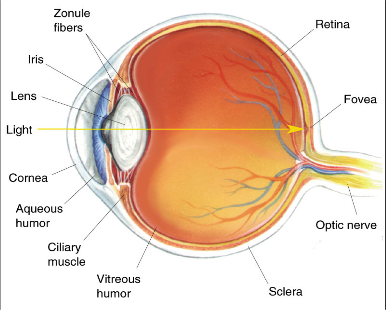
The color of the iris can be seen through the transparent cornea over it. Directly behind the iris is the lens. This structure changes shape to focus light so that we can see clearly. Its shape is convex, meaning it curves outward on both sides. The ciliary muscles above and below the lens control the shape of the lens.

Parts Of The Human Eye And Their Functions
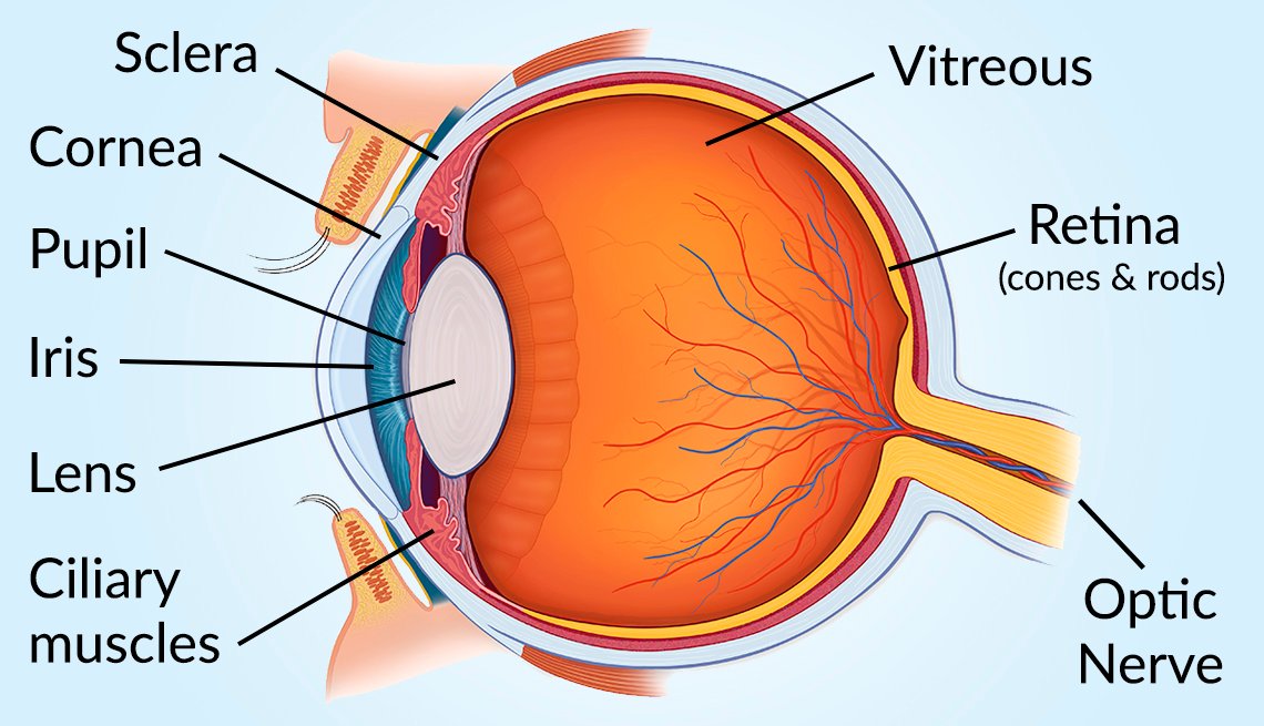
A human eye is roughly 2.3 cm in diameter and is almost a spherical ball filled with some fluid. It consists of the following parts: Sclera: It is the outer covering, a protective tough white layer called the sclera (white part of the eye). Cornea: The front transparent part of the sclera is called the cornea.

Diagram Of Human Eye Diagram Of Eye For Class 8 785x485 PNG Download PNGkit
Solution Eye: The eyes are organs that allow you to see. The eye is a sensory organ. It collects light from the visible world around us and converts it into nerve impulses. The optic nerve transmits these signals to the brain, which forms an image so thereby providing sight.
Vision and Eye Diagram How We See
The inferior rectus: Attaches to the bottom of the eye and allows downward eye movement. The medial rectus: Attaches to the side of the eye adjacent to the nose and helps the eyes to shift inwards towards the nose. The lateral rectus: Attaches to the outer side of the eyes and moves the eyes toward the temples.
Draw a labelled sketch of the human eye.
Thanks for watching our Channel. human eye diagram,human eye diagram for class 8,construction of human eye diagram,drawing human eye drawing easy step,human.
Human Eye Diagram, How The Eye Work 15 Amazing Facts of Eye
Biology Article Diagram Of Eye Diagram Of Eye The human eye is responsible for the most important function of the human body, the sense of sight. It consists of several distinct parts that work in coordination with each other. The most common eye diseases include myopia, hypermetropia, glaucoma and cataract.

Vector structure of the human eye Science notes, Medical student study, Basic anatomy and
The human eye operates similar to a digital camera in several ways: Light focuses mainly on the cornea, which acts like a camera lens. The iris controls the light that reaches the eye by adjusting the size of the pupil, and thus it functions like the diaphragm of a camera. The lens of the eye is located behind the pupil, and it focuses light.
The eye is our window to the brain and there's a lot we can tell from it
Human Eye Diagram: Contrary to popular belief, the eyes are not perfectly spherical; instead, it is made up of two separate segments fused together. Explore: Facts About The Eye To understand more in detail about our eye and how our eye functions, we need to look into the structure of the human eye. Recommended Video: 1,221
Anatomy of the Eye Human Eye Anatomy Owlcation
Labelling the eye. Use this interactive to label different parts of the human eye. Drag and drop the text labels onto the boxes next to the diagram. Selecting or hovering over a box will highlight each area in the diagram. The human eye has several structures that enable entering light energy to be converted to electrochemical energy.
eye diagram in hindi
Human Eye Diagram The figure systematically shows the components of the eye. (Image will be uploaded soon) Parts of the Human Eye The human eye has a nearly spherical shape of diameter about an inch having various parts in such a small sphere are described below: Cornea (Image will be uploaded soon)
Eye Diagram Cliparts.co
Behind the anterior chamber is the eye's iris (the colored part of the eye) and the dark hole in the middle called the pupil. Muscles in the iris dilate (widen) or constrict (narrow) the pupil to control the amount of light reaching the back of the eye. Directly behind the pupil sits the lens. The lens focuses light toward the back of the eye.
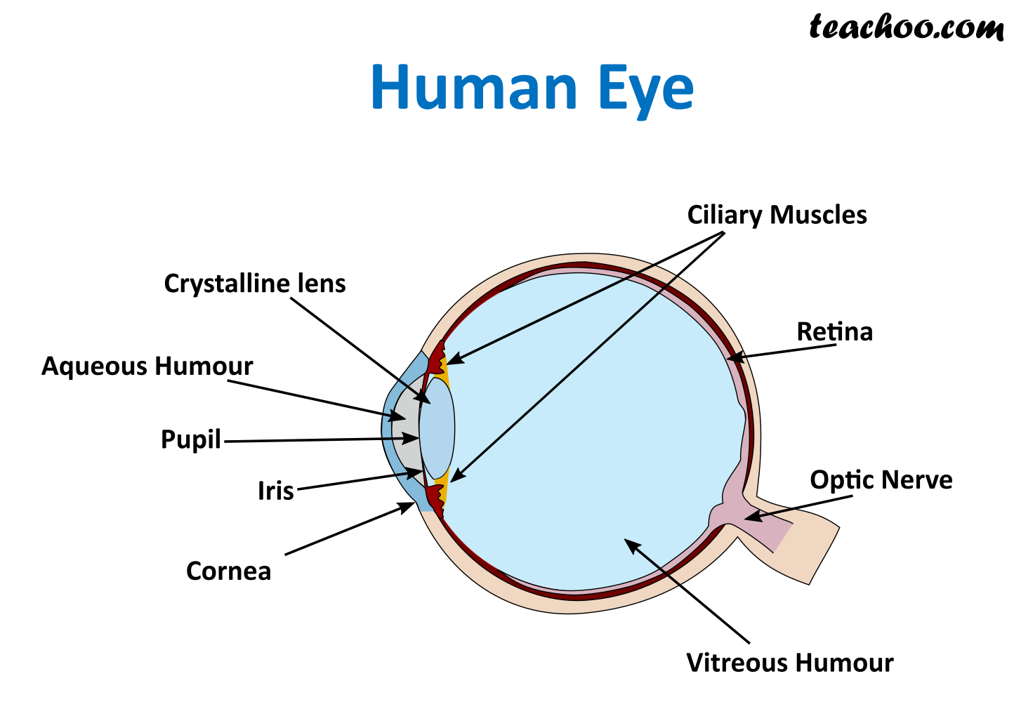
What is inside our eyes? Light Class 8 Science Teachoo
In NCERT solutions for class 8 science, students can read more about the human eye structure, function, and diagram in class 8 science chapter 16 light. Definition of the Human eye We define the human eye as the sense organ of vision. From the definition of the human eye, we understand that it is an organ that helps us to view our surroundings.

diagram of human eye with lable class 8 Brainly.in
Hi friends, In this video we will learn how to draw diagram of human eye (this diagram is based on class 8 NCERT textbook)#humameyediagram #humane.