PPT Ventricles, meninges and vessels of the CNS PowerPoint
Definition Die Hirnventrikel sind ausgedehnte, mit Liquor gefüllte Hohlräume im Inneren des Gehirns, die durch Foramina und Verbindungsstrukturen (beispielsweise den Aquaeductus mesencephali) miteinander kommunizieren. Anatomie
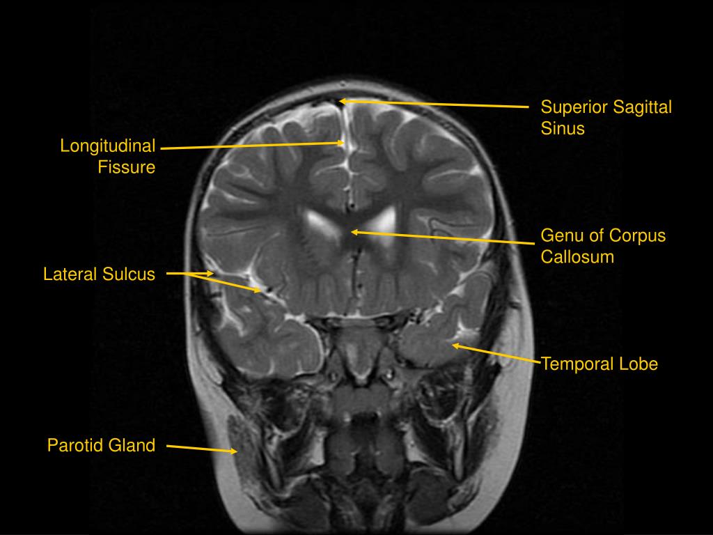
PPT MRI of Brain/Head and Neck PowerPoint Presentation, free download
Cornu temporale. Definition. There is no definition for this structure yet. Suggest a definition I agree herein to the cession of rights to my contribution in accordance with the Terms and conditions of the website. Cancel Submit. I agree herein.
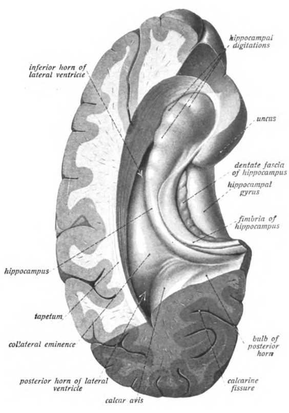
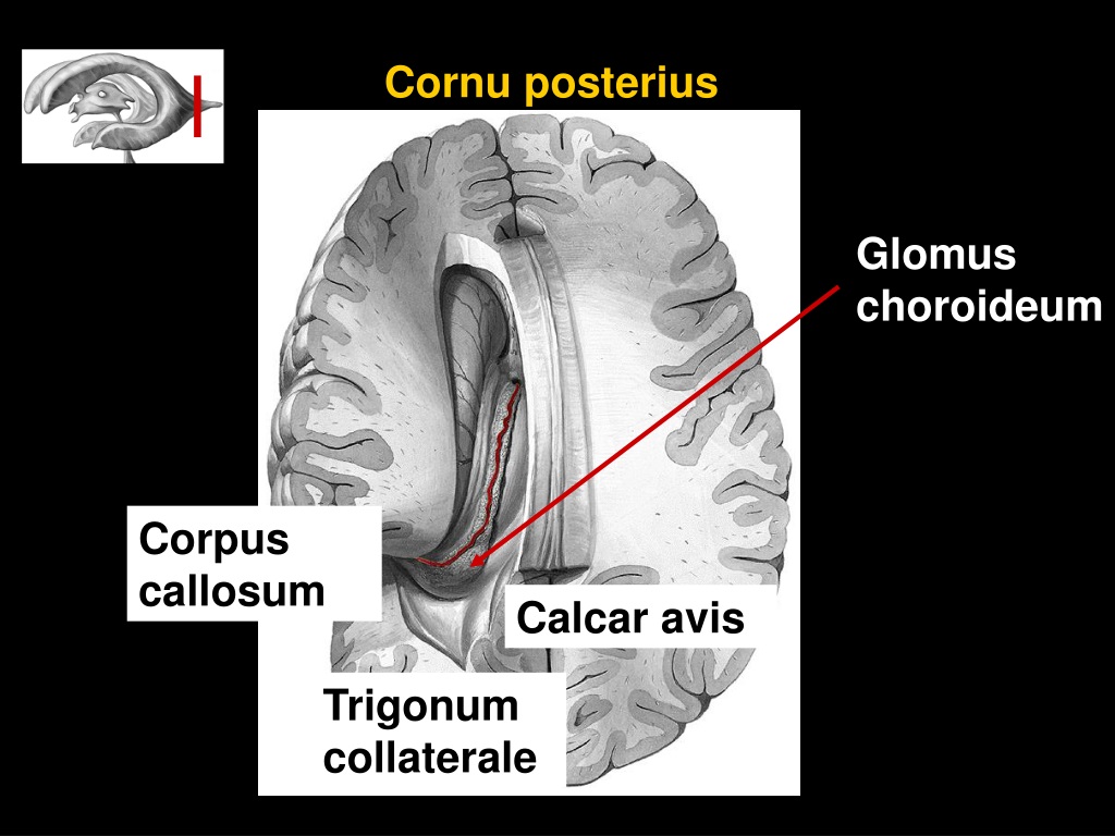
Sobotta 1909 fig.639 Posterior and inferior horns of the lateral
Ncl. ventralis anterior thalami. ventral anterior thalamic nucleus. Tr. amygdalofugalis ventralis. ventral anygdalofugal pathway. Authors & Publisher. Brain in the Head. The consistent and unified anatomical terminology of the Nomenclature is the basis for the Atlas of the Human Brain and all supplemental material.
Temporal Horn Diagram Free Download Wiring Diagram Schematic
The AD patients have more remarkable atrophy of entorhinal cortex, perirhinal cortex, and have obvious extension of cornu temporale and uncus distance in comparison with the normal controls. The shrinkage rate of hippocampus can be used as a marker for the diagnosis and progress of AD.
Temporal bone Anatomy, parts, sutures and foramina Kenhub
From 'Atlas and Textbook of Human Anatomy', 1909, Vol. 3, fig.674, by Johannes Sobotta and J. Playfair McMurrich. Artist: K. Hajek. Retrieved from Sobotta's Anatomy plates at Wikimedia. Possible original source: Sobotta's atlas at Hathitrust Digital library. Creator (s)/credit: Prof.dr. Johannes Sobotta, anatomist. You are free to use this item.
Ventricles and Coverings of the Brain Neupsy Key
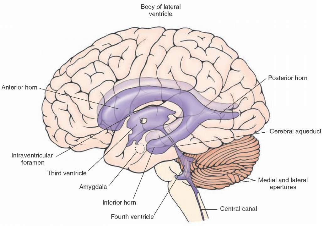
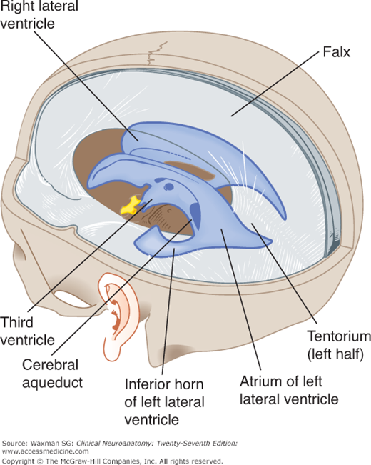
Description: In this 3D model, the ventricles of the brain and their adjecent structures are shown. Anatomical structures in item: Cornu frontale ventriculi lateralis Cornu occipitale ventriculi lateralis Cornu temporale ventriculi lateralis Ventriculus quartus Apertura lateralis ventriculi quarti Aqueductus mesencephali Foramen interventriculare

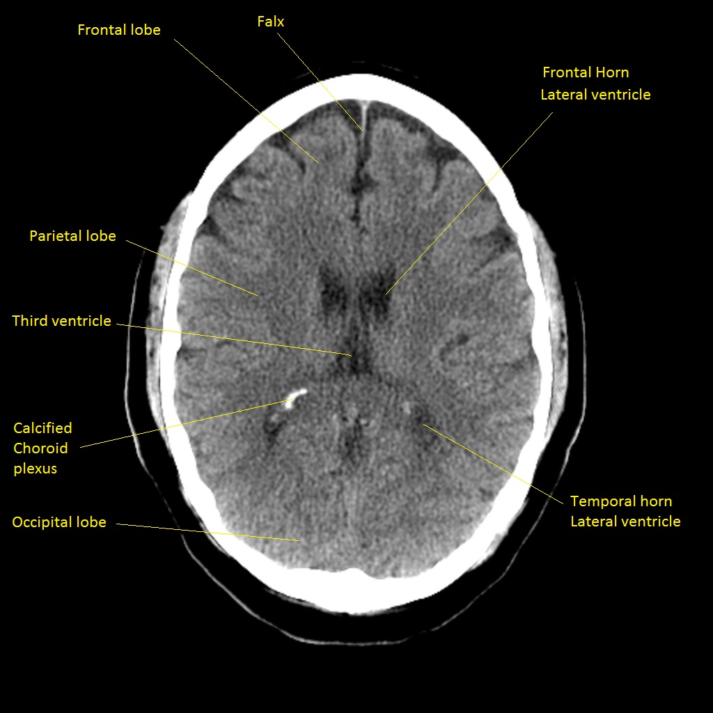
Pin by Dr abuaiad on brain&head and neck Radiology, Brain diagram
Anatomy of the ventricular system. The ventricular system forms the cavities of the central nervous system and is filled with cerebrospinal fluid.
Coronal Section Through the Cerebrum ClipArt ETC
sea turtle fact sheet, 2005. they reach sexual maturity and are ready to. A rehabilitated sea turtle makes its. mate. Although sea turtles can live to be over way back to the ocean. 50 years old, they have a very low survival rate. Only about one in 1, hatchlings will 000 live to reproduce.
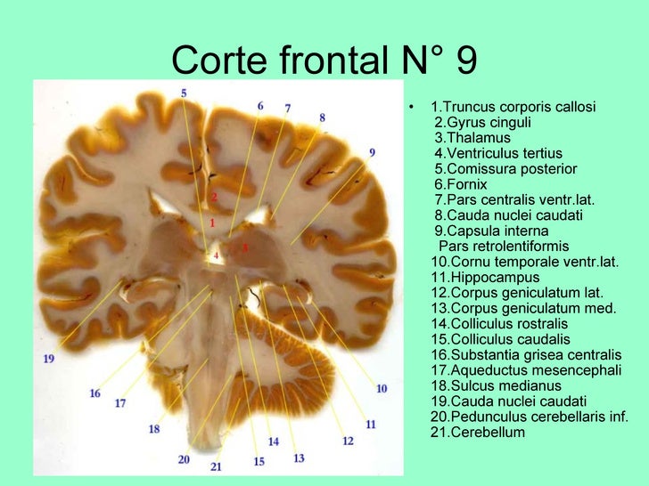
corte coronal de encefalo
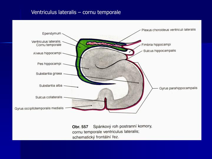
Cornu temporale ventriculi lateralis Quick Facts The lateral ventricles can be divided into three horns; the frontal, temporal and occipital horns. The temporal horn of lateral ventricle (aka inferior horn of lateral ventricle) is located inferior to the atrium and extends antero-inferiorly towards the temporal lobe. Complete Anatomy

Log On to Constellation Hemisphere, Cerebral cortex, Corpus callosum
Ventriculus lateralis, Cornu temporale Capsula interna Nucleus caudatus (font: arial black, size: 10) Date 30 November 2005 Source http://www.healcentral.org/healapp/showMetadata?metadataId=40566(Internet Archive of file description page)
PPT THE LIMBIC SYSTEM PowerPoint Presentation ID491441
. The hippocampus is found deep within the medial temporal lobe of the brain, being part of the limbic system. Its main roles include consolidation of declarative (episodic) memories and.
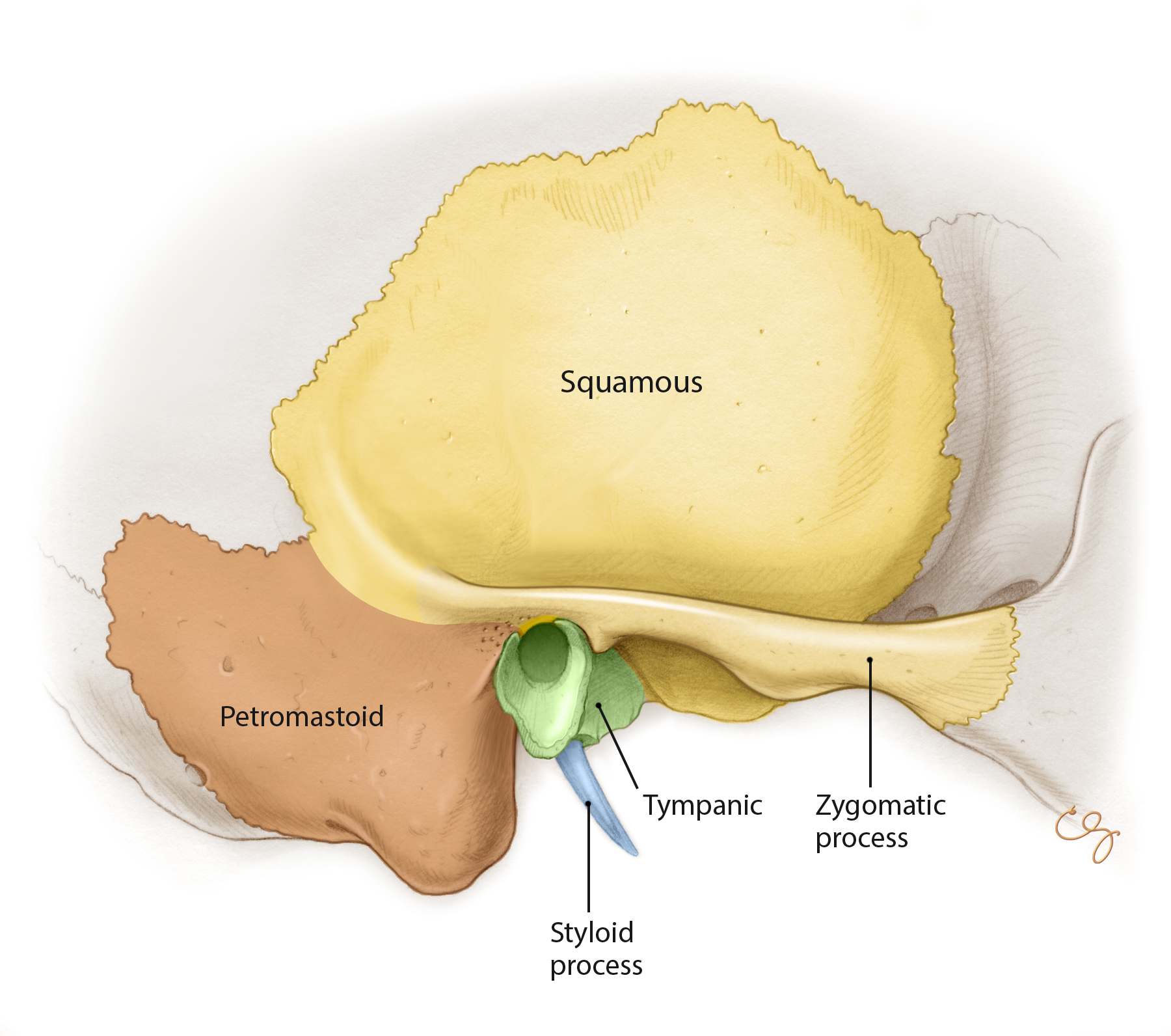
Overview of Temporal Bone Oto Surgery Atlas
Antenatal choroid plexus cysts. Antenatal choroid plexus cysts are benign cysts that occur due to an infolding of the neuroepithelium in a fetus, most commonly in the lateral ventricles. Ranging from a few mm to 1-2cm in size, these cysts generally occur in approximately 2% of all pregnancies.

Hirnventrikel (Vorschau) Anatomie des Menschen Kenhub YouTube
the part of the lateral ventricle extending downward and forward into the medial part of the temporal lobe. See: lateral ventricle.
Hipocampo Anatomia, funções e conexões Kenhub
The fluid (cerebrospinal fluid) is produced in the ventricular system of the brain. There are four such hollow spaces in the brain that house cerebrospinal fluid (CSF): two lateral ventricles, a third ventricle and a fourth ventricle. This article will look at the structure of this system and how it helps the brain. Contents Choroid plexus

Lobes of the Brain Cerebral Cortex Anatomy, Function, Labeled Diagram
At certain sites e.g. on the cornu ammonis, suprachiasmatic and infundibular recesses, on the bottom of the aquaeductus mesencephali small foci without microvilli and cilia ("bare") can be observed. In the upper half of the suprachiasmatic region some of the cilia have button-like and in the cornu temporale in the lateral ventricle club-like.
ABC Medical Comprehensive Medical Encyclopedia Education
You are free: to share - to copy, distribute and transmit the work; to remix - to adapt the work; Under the following conditions: attribution - You must give appropriate credit, provide a link to the license, and indicate if changes were made. You may do so in any reasonable manner, but not in any way that suggests the licensor endorses you or your use.