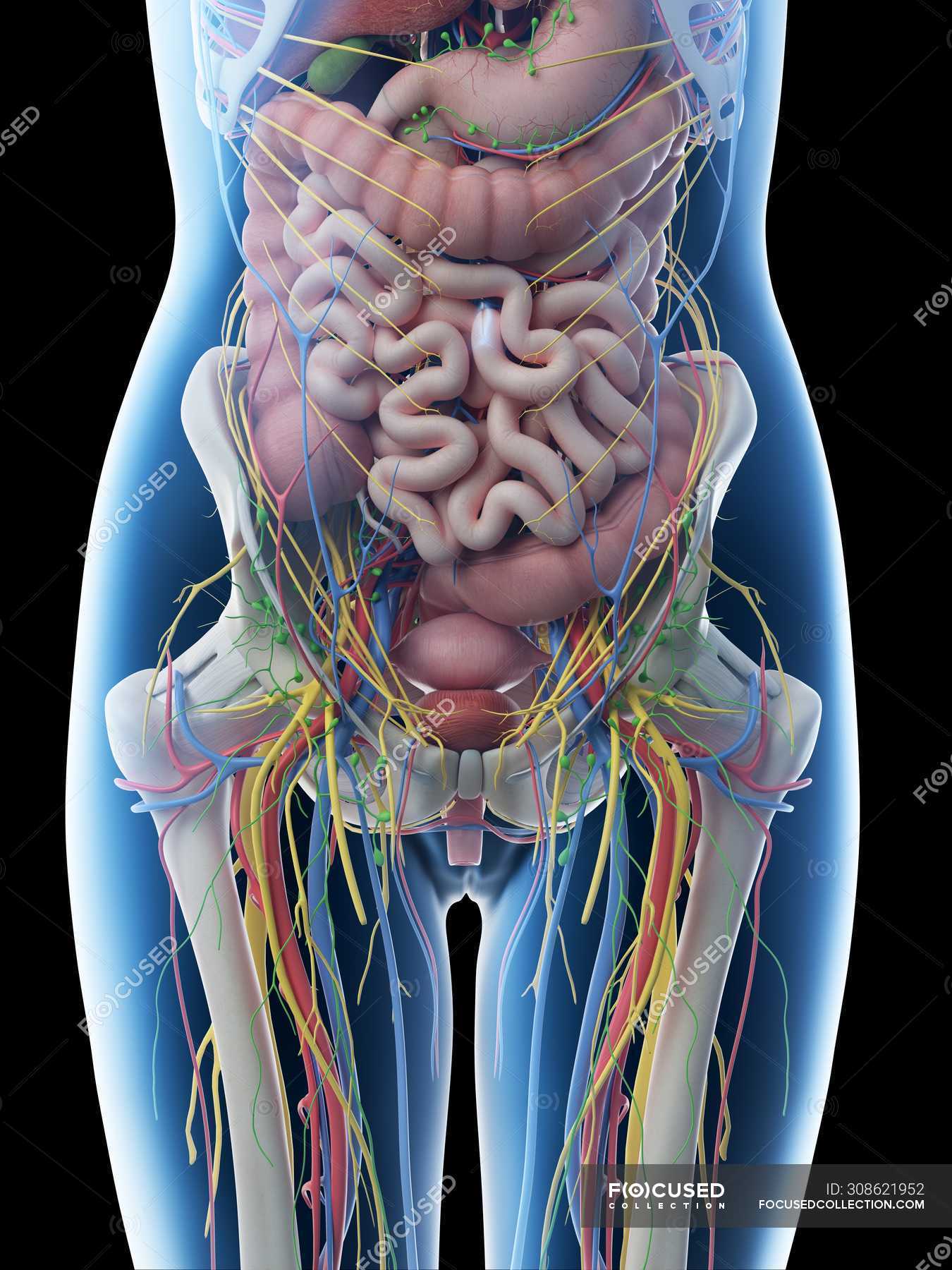
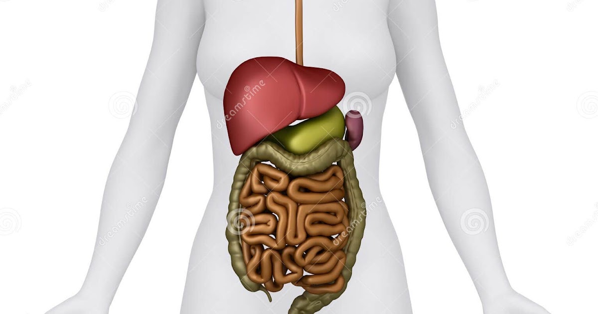
Female abdominal anatomy and internal organs, computer illustration
Uterus. Also called the womb, the uterus is a hollow, pear-shaped organ located in a woman's lower abdomen, between the bladder and the rectum. Ovaries. Two female reproductive organs located in the pelvis. Fallopian tubes. Carry eggs from the ovaries to the uterus. Cervix.

Abdomen Wikipedia, la enciclopedia libre
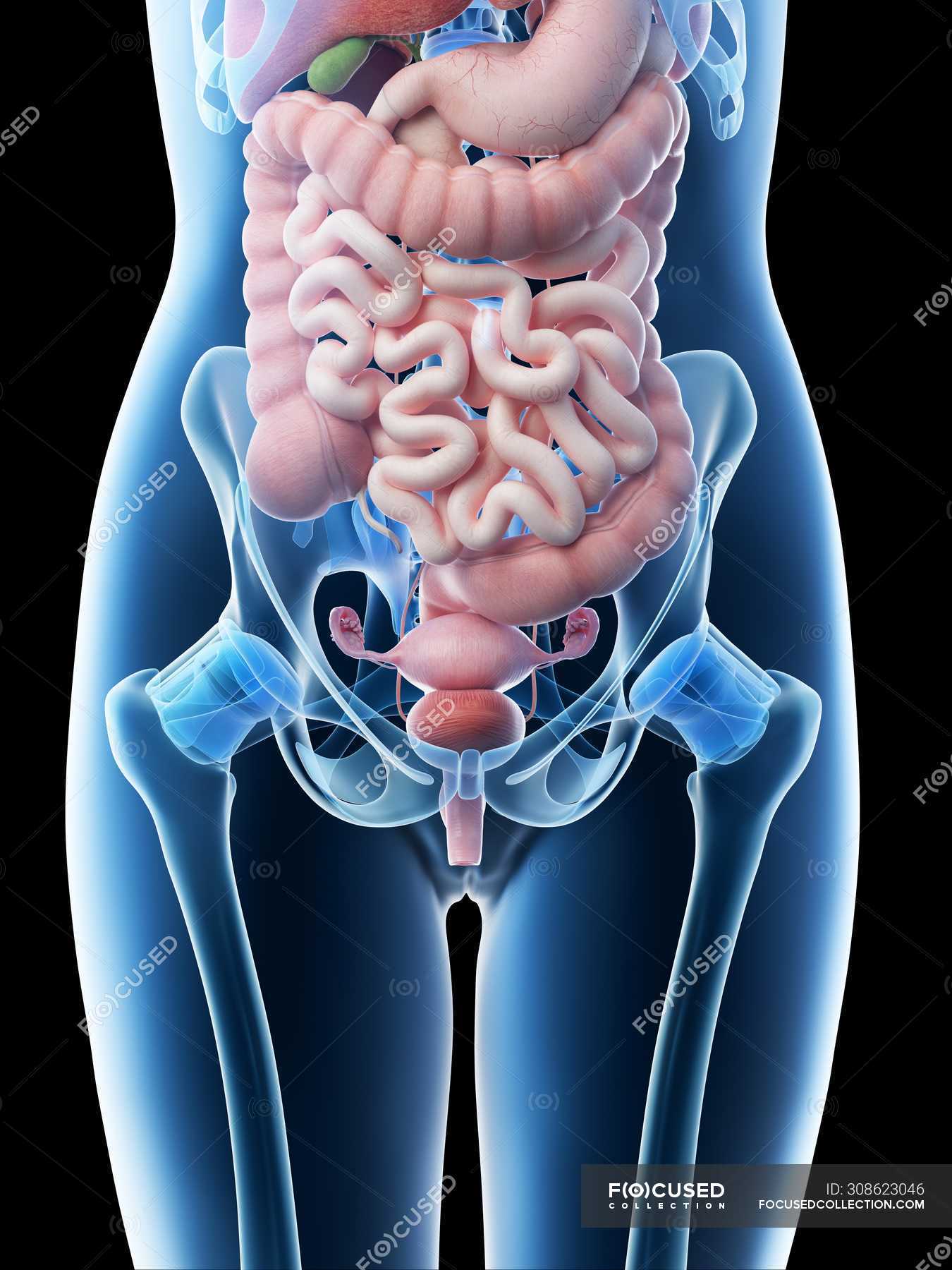
Browse Anatomy of the Female Abdomen and Pelvis ID: exh6130a Cite this Item Add to Collection This medical illustration depicts a mid-sagittal view of the normal anatomy of the female abdomen and pelvis. Labeled structures include the large bowel (colon or large intestine), umbilicus, small intestine, ovary, fallopian tube, uterus and bladder.

Female Anatomy Upper Body Stock Photo Download Image Now iStock
Browse 2,691 female stomach anatomy photos and images available, or start a new search to explore more photos and images. NEXT Browse Getty Images' premium collection of high-quality, authentic Female Stomach Anatomy stock photos, royalty-free images, and pictures.
Female Abdominal Anatomy Pictures / Stock Images Female Abdominal
Sources + Show all Anterolateral abdominal wall Surface anatomy Let's first take a look at the surface anatomy of the anterolateral abdominal wall, before we dive into its layer description. The anterolateral abdominal wall spans the anterior and lateral sides of the abdomen.
Abdominal Anatomy Anatomy Of The Female Abdomen And Pelvis Cut Away Images
The retroperitoneal structures include the suprarenal glands, aorta and inferior vena cava, duodenum (parts 2 to 4), pancreas (head and body), ureters, colon (descending and ascending), kidneys, esophagus (thoracic), and rectum. The abdomen derives from three primary germ layers as an embryo. These are the ectoderm, which forms the epidermis.

Female Anatomy Stock Photo Download Image Now Abdomen, Anatomy
The abdomen (colloquially called the belly, tummy, midriff, tucky or stomach) is the part of the body between the thorax (chest) and pelvis, in humans and in other vertebrates. The abdomen is the front part of the abdominal segment of the torso. The area occupied by the abdomen is called the abdominal cavity.
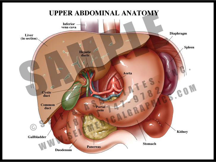
Upper Abdominal Anatomy S&A Medical Graphics
Anatomy atlas of the female pelvis: 101 labeled illustrations of the female genital system (ovaries, uterine tubes, uterus, vagina, vulva, clitoris) and pelvic cavity (bladder, rectum, pelvic diaphragm, perineum with innervation and blood supply). Tome 2 : Thorax, coeur, abdomen et pelvis. Torsten B. Möller - Emil Reif. Paru le : 06/2014.

Female Abdominal Anatomy TrialExhibits Inc.
The abdomen is the part of the body that contains all of the structures between the thorax (chest) and the pelvis, and is separated from the thorax via the diaphragm.. In this section, learn more about the anatomy of the abdomen- its areas, bones, muscles, the gastrointestinal tract, accessory organs and the abdominal vasculature. Areas of.
Abdomen AnatomyFemale Female Abdominal Anatomy Illustration Stock
The pelvic floor is a unique anatomical location where the balance of the different pressures, either visceral, muscular, or liquid play a fundamental role in the physiological functioning of all the structures contained therein. The pelvis is bounded superiorly by the imaginary line between the pubis and sacral promontory and inferiorly as the line between the ischial tuberosity and the apex.
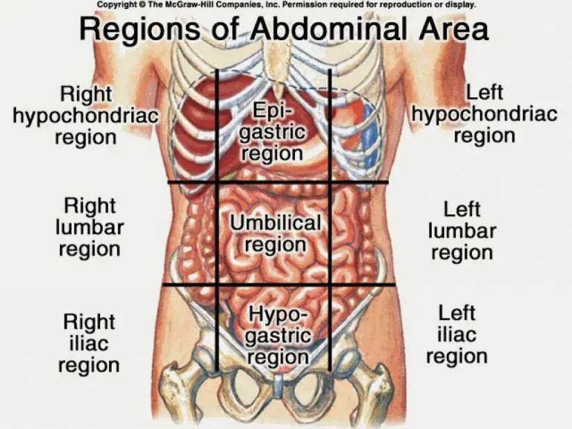
Female Abdomen Anatomy Quadrants / Abdominal Surface Anatomy Radiology
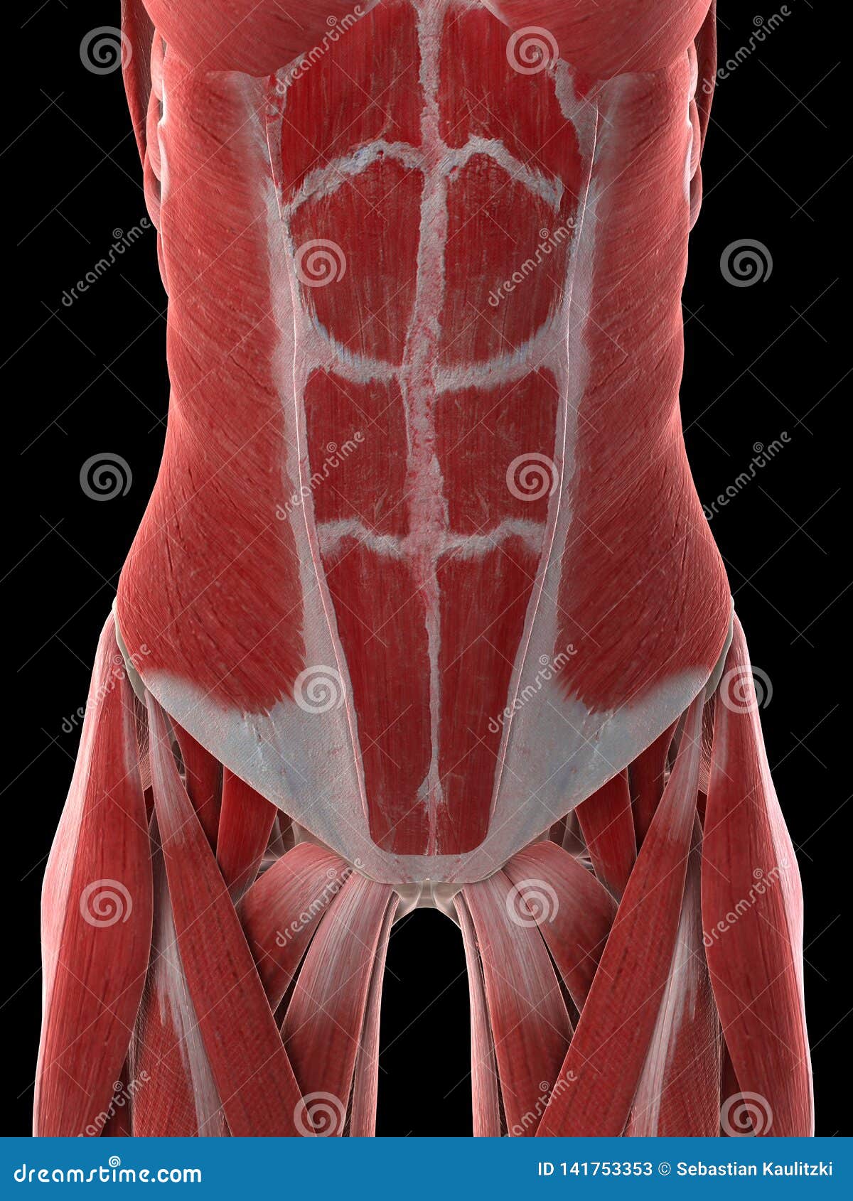
Abdomen The muscles of the abdomen protect vital organs underneath and provide structure for the spine. These muscles help the body bend at the waist. The major muscles of the abdomen include.
Abdomen Anatomy Female Body Illustration of female digestive system
Fact checked by Sarah Scott Table of Contents View All Diagram External Internal Breast Anatomy Functions Female anatomy includes the internal and external structures of the reproductive and urinary systems. Reproductive anatomy plays a role in sexual pleasure, getting pregnant, and breastfeeding.
A Females Abdominal Muscles Stock Illustration Illustration of muscle
The abdomen describes a portion of the trunk connecting the thorax and pelvis. An abdominal wall formed of skin, fascia, and muscle encases the abdominal cavity and viscera. The abdominal wall does not only contain and protect the intra-abdominal organs but can distend, generate intrabdominal pressure, and move the vertebral column. Detailed knowledge of the components of the abdominal wall is.

Human Anatomy Female Abdomen Peritoneum And Peritoneal Cavity Anatomy
Overview What are the abdominal muscles? Your abdominal muscles are a set of strong bands of muscles lining the walls of your abdomen (trunk of your body). They're located toward the front of your body, between your ribs and your pelvis. There are five main muscles in the abdomen: External obliques. Internal obliques. Pyramidalis. Rectus abdominis.
Anatomy Of The Female Abdomen And Pelvis, Cut away View Healthiack
The levator ani muscles consist of three separate muscles: Puborectalis. This muscle is responsible for holding in urine and feces. It relaxes when you urinate or have a bowel movement.

Female Abdominal Organs Diagram Human anatomy woman abdomen Humas
Show details Anatomy, Abdomen and Pelvis: Female Pelvic Cavity Austin McEvoy; Maggie Tetrokalashvili. Author Information and Affiliations Last Update: July 24, 2023. Go to: Introduction The pelvic cavity is a bowl-like structure that sits below the abdominal cavity. The true pelvis, or lesser pelvis, lies below the pelvic brim (Figure 1).

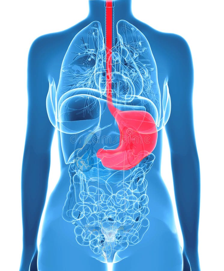
Anatomy of female stomach, illustration Stock Image F010/9291
Reading time: 17 minutes Recommended video: Surface anatomy of the abdomen and the lower extremity [13:14] Overview of the surface anatomy landmarks found in the abdomen and lower limbs. Abdomen 1/2 Synonyms: Abdominal region, Regio abdominis , show more. Hello there fellow anatomist and welcome to abdomen and pelvis 101!